
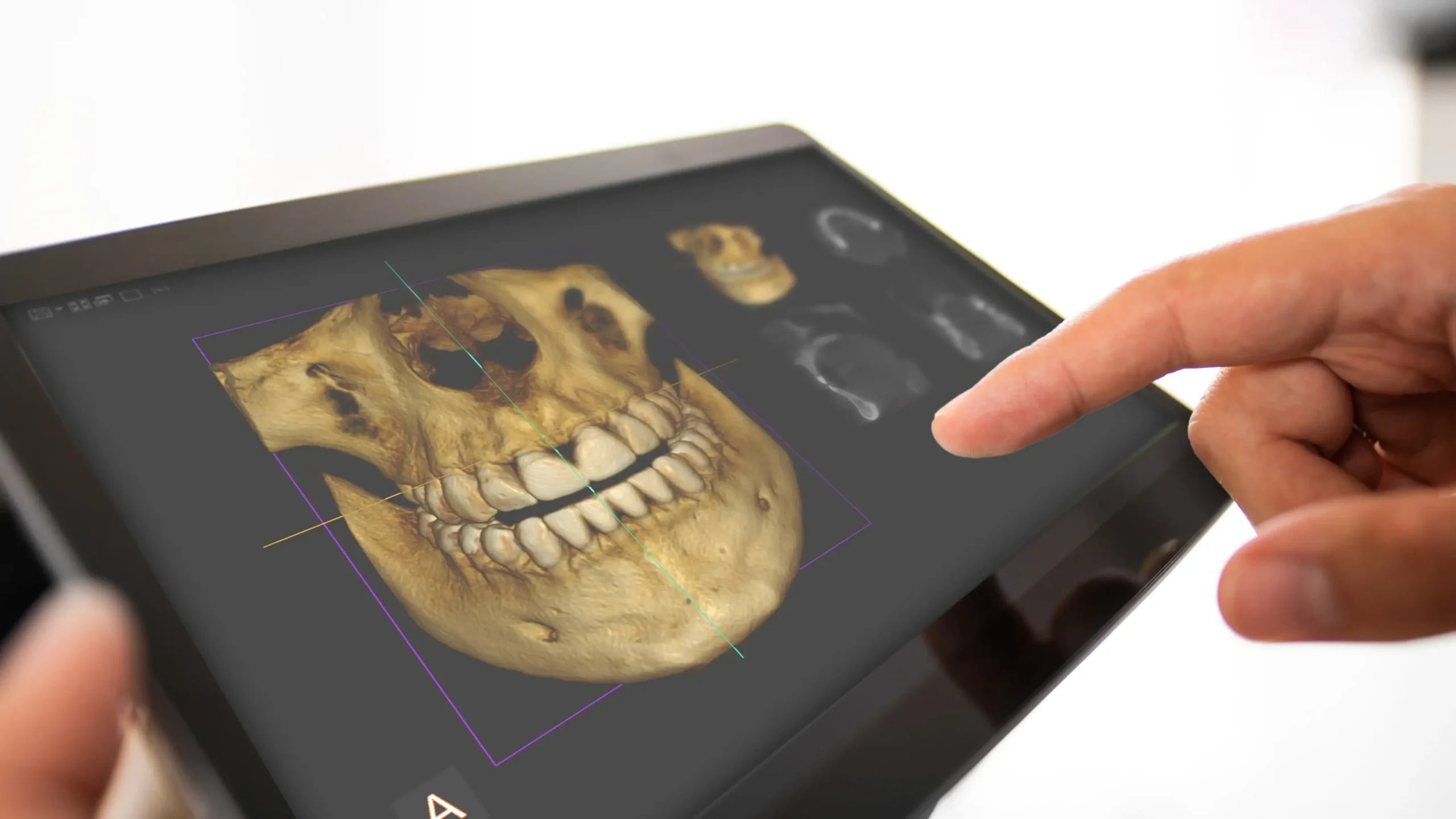
3D Dental Imaging Technology: Diagnostic Excellence at Dentocare Kahramanmaraş
Imaging technologies, which have a revolutionary place among medical diagnostic methods, also undertake the mission of “making the invisible visible” in dentistry. In complex cases where traditional methods are insufficient, the three-dimensional examination of anatomical structures is the most critical element determining the fate of the treatment. Dentocare, aiming for technological leadership in the field of oral and dental health in the Kahramanmaraş region, offers its patients the most accurate diagnosis and the most reliable treatment planning with its advanced 3D Dental Imaging systems. This technology, also known as Dental Volumetric Tomography (CBCT), allows for the analysis of jawbone, teeth, nerve pathways, and soft tissues with millimeter precision.
What is 3D Dental Imaging?
3D Dental Imaging, or Cone Beam Computed Tomography (CBCT) as it is technically called, is an X-ray technology specifically developed for dentistry. Unlike classic medical CT scans, it uses a conical X-ray beam. This system takes hundreds of cross-sections of the patient’s head and neck region with a single scan and combines this data through powerful computer software to create a 3D model. On this model, bone density, the angle of tooth roots, and anatomical spaces can be examined from every angle. This system, used at Dentocare Kahramanmaraş clinic, forms the cornerstone of surgical planning.
How Does 3D Dental Imaging Technology Work?
The device rotates 360 degrees (or partially) around the patient’s head while standing or sitting. During this rotation, the X-ray source and detector move synchronously. The cone beam scans the target area, and the raw data obtained is transferred to a digital environment. Thanks to three-dimensional pixels called “voxels,” the image can be rotated in any direction, cross-sections can be taken, and magnification can be performed without compromising image quality. Unlike traditional X-rays, the images do not overlap; each structure is clearly seen in its own spatial position.
The Importance of 3D Imaging in Dentistry
In dentistry, accurate diagnosis is half the battle. In two-dimensional images (panoramic or periapical X-rays), there is no perception of depth, and anatomical structures can be shadowed by each other. 3D imaging, on the other hand, allows the doctor to virtually tour the inside of the patient’s jaw structure. Especially in risky procedures such as implant surgery, impacted tooth extractions, and cyst operations, 3D imaging is vital to minimize the risk of nerve damage and shorten the operation time.
Differences Between 3D Dental Imaging and 2D X-rays
Understanding the differences between traditional methods and modern 3D technology explains why tomography is necessary in some cases. Here are the key differences:
| Features | 2D X-ray (Panoramic/Periapical) | 3D Dental Imaging (CBCT) |
|---|---|---|
| Size Perception | Height and Width Only (Planar) | Height, Width and Depth (Volumetric) |
| Magnification/Distortion | Image magnification/distortion may occur by 10-25% | One-to-one (1:1) measurement capability, zero deterioration |
| Details | Structures overlap (Superposition) | Each structure is viewed separately and clearly |
| Area of Use | General inspection, caries detection | Implant, cyst, impacted tooth, trauma |
| Radiation Dose | Very low | Low (90% less than medical CT) |
Advantages of 3D Dental Imaging
3D imaging protocols applied at Dentocare Kahramanmaraş, both It provides numerous advantages to both the doctor and the patient:
- Accurate Diagnosis: Clarifies suspicious situations and eliminates the question of “what if”.
- Safe Surgery: The exact location of critical structures such as the mandibular nerve (mandibular canal) and sinus cavities is determined.
- Shorter Treatment Time: The procedure is completed faster because there are no surprises during surgery.
- Patient Communication: The patient understands their treatment needs better by seeing their jaw structure in 3D.
- Minimally Invasive Procedure: Since the bone condition is seen before making an incision, the procedure can be performed with smaller incisions (flapless surgery).
Detailed Oral and Jaw Analysis with 3D Imaging
This technology analyzes not only the teeth but also the surrounding tissues. The quality (density), width, and height of the jawbone are measured. Pathologies in the sinus cavities (sinusitis, polyps, etc.), deformities in the temporomandibular joint (TMJ), and airway problems are also examined.Narrowings in the jaw can be detected. Thus, the dentist can evaluate the patient’s craniofacial health holistically, not just limited to the oral cavity.
Which Treatments Use 3D Dental Imaging?
3D tomography is no longer a luxury diagnostic tool, but a standard requirement for many treatments. Its uses include:
3D Dental Imaging in Implant Treatments
Implant success depends on the correct bone volume and placement at the correct angle. With 3D imaging, bone thickness is measured in millimeters. The dentist virtually places the implant into the bone and practices it. In this way, the risk of hitting a nerve or drilling through the bone during the operation is eliminated. At Dentocare in Kahramanmaraş, implant treatments are carried out with 100% digital planning.
3D Imaging in Jaw and Joint Assessment
Temporomandibular joint disorders are difficult to diagnose. In patients experiencing pain, clicking sounds, or limited mouth opening, the condition of the condyle and joint socket is visualized in 3D. Problems such as calcification, wear, or displacement are clearly identified, allowing for the creation of an accurate treatment plan.
3D Imaging in the Evaluation of Impacted Teeth
Impacted wisdom teeth, in particular, can sometimes be very close to nerve canals or have hook-shaped roots. This proximity is not fully understood in 2D X-rays. 3D tomography shows precisely whether the tooth roots are in contact with the nerve and in which direction the tooth is tilted. This allows the surgeon to perform the tooth extraction with minimal trauma.
3D Dental Imaging in Root Canal Treatments
Persistent pain or failed root canals are often caused by undetected extra canals or root tip infections. 3D imaging maps the internal structure of the tooth. The boundaries of overlooked canals, root cracks, or cysts become clearer. This makes it possible to save the tooth instead of extracting it (Endodontic Microsurgery).
3D Imaging in Orthodontic Treatment Planning
In braces or clear aligner applications, the position of the tooth roots within the bone is important. Also, if the eruption of impacted canine teeth is planned, the exact position of the tooth is determined with 3D. 3D analysis is mandatory in skeletal disorders requiring orthognathic surgery (jaw surgery).
3D Dental Imaging Before Surgical Procedures
Before cyst or tumor operations, bone grafting (bone powder) applications, and sinus lifting procedures, a volumetric analysis of the surgical field is performed. The boundaries of the pathology are determined, and the removal of the lesion is planned without damaging the surrounding tissues.
Precise Treatment Planning with 3D Dental Imaging
Planning is the mirror of the application. Dentocare dentists design surgical guides by processing 3D data in specialized software. These guides are templates that ensure the implant is placed precisely at the planned location. Precise planning minimizes complications such as postoperative swelling, pain, and bruising.
How Long Does a 3D Dental Imaging Procedure Take?
The speed of technology directly affects patient comfort. The 3D scanning process is extremely fast. It takes an average of 10 to 20 seconds for the device to rotate around the patient and acquire the image. Processing the image on the computer and displaying it on the screen takes a few minutes. Therefore, all diagnostic data is obtained in a total of 5 minutes.
Is 3D Dental Imaging Safe?
Radiation dose is one of the most frequently asked questions by our patients. Dental CBCT devices emit much lower radiation than general body CT scans (Medical CT) taken in hospitals (approximately 10-15 times less). Thanks to the sensitivity of digital sensors, the necessary information is obtained with a minimum dose. Operating on the “ALARA” (Lowest Possible Dose) principle, Dentocare prioritizes patient safety above all else.
Who is 3D Dental Imaging Suitable For?
It is suitable for patients of all ages requiring detailed diagnosis, except pregnant women (except in emergencies and under doctor’s supervision). It is ideal especially for those who will have implants, those with impacted teeth, those who have experienced trauma, and those with unexplained toothaches.
3D Dental Imaging in Children and Adults
It can be safely used in children to detect dental development disorders, cleft palates, or impacted teeth. Special low-dose modes (Pediatric Mode) are available for children. In adults, it is frequently used for bone loss detection and prosthesis planning. Thanks to the open design of the device, patients and children with claustrophobia can have the imaging done comfortably.
Early Diagnosis with 3D Dental Imaging
Some intraosseous lesions or cysts can grow without showing any signs on 2D X-rays and may only be detected when they are very advanced. 3D imaging detects even the slightest changes in bone density, allowing problems to be resolved before they grow. This is preventive.3D imaging is the most powerful weapon in dentistry.
Digital Archiving and 3D Image Recordings
The data obtained is entirely digital. This ensures that patient records are securely stored, can be shared with other specialists (consultation) when needed, and allow for comparison of changes over the years. The need for physical film storage is eliminated.
Studies show that the success rate of treatments planned with 3D imaging is significantly higher than with traditional methods. Accurate measurement, correct material selection, and the correct surgical approach prevent implant loss and treatment repetitions.
Immediately after the imaging, the images appear on the dentist’s screen. The dentist examines the images with the patient. They show pathologies on the cross-sections, measure bone thickness, and explain the treatment plan. The patient, seeing their own jaw structure in 3D, consciously approves the treatment.
Modern Technological Infrastructure in 3D Dental Imaging
Dentocare Kahramanmaraş boasts one of the most advanced imaging centers in the region. The equipment is equipped with high-resolution (HD) mode, artifact reduction technology, and fast scanning capabilities. This technological infrastructure guarantees the accuracy of the diagnosis.
Predictable Treatment Outcomes with 3D Dental Imaging
In dentistry, “surprises” are undesirable. 3D technology makes the outcome of treatment predictable. Where the implant will be placed, how the tooth will be extracted, and how far the root canal will be filled are all known in advance. This predictability reduces stress for the dentist and increases patient confidence.
Safer Dental Treatments with 3D Dental Imaging
Your health is too valuable to leave to chance. 3D Dental Imaging is one of the greatest privileges offered by modern medicine, and at Dentocare Kahramanmaraş, this privilege is a standard service. Introducing the technology that unlocks the secrets deep within your jawbone, plans your treatment with mathematical precision, and maximizes your safety. Accurate diagnosis is the key to successful treatment; and we use that key for you.