
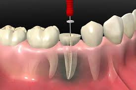
Root Canal Treatment with Microscope: Success Hidden in the Details with Dentocare Kahramanmaraş
Dentistry is a delicate science requiring work in areas as small as one-tenth of a millimeter. Especially in treatments performed in complex, narrow, and dark systems such as the root canals of a tooth, the dentist’s vision is the most critical key to success. In root canal treatments performed with traditional methods, dentists usually worked with the naked eye or with magnifying glasses (loupes), and microscopic details that could be missed could affect the long-term success of the treatment. However, with the beginning of the “Micro-Endodontics” era, dental microscopes have completely changed the course of treatment. Dentocare, a pioneer of technological dentistry in Kahramanmaraş, manages to save even teeth that were previously decided to be extracted by shedding light on the deepest points of the tooth with microscope-assisted root canal treatment.
What is Root Canal Treatment with a Microscope?
Root canal treatment with a microscope is an advanced endodontic procedure that magnifies the internal structure of the tooth up to 25 times, allowing for crystal-clear visualization of details invisible to the naked eye. This method eliminates the dentist’s reliance on guesswork based solely on touch and X-ray images; instead, it offers the possibility of treating “seeing and knowing.” A dental microscope consists of a powerful light source and high-resolution lenses. This system clearly reveals the entrances to root canals, cracks, constrictions, and anatomical variations. At Dentocare Kahramanmaraş clinic, this technology is used as a standard part of root canal treatments, maximizing success rates.
Using a Dental Microscope in Root Canal Treatment
The dental microscope is unique because, in addition to magnifying the dentist’s working field, it transmits light to the deepest point of the root canal (apex). Under normal clinical lighting, the inside of the canal is like a dark tunnel. However, the microscope, with its coaxial illumination system, directs light in the same direction as the field of view, thus preventing shadow formation. This allows the surgeon to clearly distinguish infected tissue, broken instrument fragments, or calcified areas within the canal, and to intervene only in the problematic area without damaging surrounding tissues.
Advantages of Microscope-Assisted Root Canal Treatment
The use of a microscope provides numerous advantages that increase the quality and predictability of treatment:
- Superior Visual Control: Allows for the detection of details that are not visible to the naked eye.
- Tissue-Preserving Approach: Only the infected area is cleaned, maximizing the preservation of healthy tooth tissue.
- Locating Hidden Canals: Missed canals (such as MB2), which are the main cause of treatment failure, are easily found.
- Removing Broken Instruments: Allows for the safe removal of broken instruments within the canal.
- Perforation Repair: Enables precise repair of perforations that occur at the root base.
- Ergonomics and Comfort: A more comfortable and faster process for both the doctor and the patient. presents.
Differences Between Traditional Root Canal Treatment and Microscope-Assisted Treatment
The difference between traditional methods and microscope-assisted methods can be likened to the difference between driving a car in the dark at night and driving a car in the daylight. The following table outlines the key differences between these two approaches:
| Features | Traditional Root Canal Treatment | Microscopic Root Canal Treatment |
|---|---|---|
| Magnification | None or Limited (2.5x – 4x with loupe) | High Magnification (3x – 25x) |
| Lighting | Unit light (Shadow may occur) | Coaxial LED/Xenon Light (Shadow-free, focused inside the canal) |
| Canal Detection | With the aid of touch sensation and X-ray | Direct visual detection |
| Tissue Preservation | Entry cavity can be opened wider | Minimally invasive, pinpoint entry |
| Success Rate | High (85-90%) | Very High (95-98%) |
Wide Field of View in Microscopic Root Canal Treatment
As magnification power increases, the physician’s field of view narrows, but detail control increases. Dental microscopes offer flexibility to the dentist thanks to different magnification levels (magnification changers). Low magnification is used for a general view, while high magnification is used to examine a detail at the root tip. The microscopes used at Dentocare in Kahramanmaraş allow the dentist to change focus without using their hands thanks to motorized focusing systems, which shortens the procedure time.
Detection of Hidden and Additional Root CanalsUsing a Microscope in Root Canal Treatment
Especially in upper molars, there is a probability of over 90% presence of a fourth canal called “MB2”. However, this canal is very narrow and its entrance is calcified, making it almost impossible to see with the naked eye. In traditional treatments, this canal is often overlooked and causes pain months after treatment. Under a microscope, however, these hidden canals are easily detected and cleaned with the help of special dyes and ultrasonic tips. This is the most effective way to prevent failed root canal treatments (retreatment).
In Which Cases is Microscopic Root Canal Treatment Preferred?
Actually, every root canal treatment deserves to be performed under a microscope, but in some cases its use becomes a necessity:
- In the renewal of previously performed but unsuccessful root canal treatments (Retreatment).
- In teeth with calcification or blockage in the root canals.
- In cases where the root canal instrument breaks.
- In cases requiring root tip surgery (Apical Resection).
- In the treatment of young teeth that have not completed root development (Apexification).
- In the diagnosis of cracks in the tooth that are not visible to the naked eye.
Examination and Planning Before Microscopic Root Canal Treatment
At Dentocare Kahramanmaraş clinic, the process begins with a detailed radiological examination. In addition to periapical X-rays, a three-dimensional anatomy of the tooth is obtained using 3D Dental Tomography (CBCT). The microscope allows for the verification of this radiological data in a clinical setting. The dentist shapes the treatment plan by seeing the complex structure seen in the tomography under the microscope.
How is Root Canal Treatment Performed Under a Microscope?
The procedure follows standard root canal treatment procedures, but each stage is performed under a microscope:
- Anesthesia and Isolation: The tooth is numbed and isolated with a rubber dam.
- Entry Cavity: The inside of the tooth is accessed under a microscope with minimal loss of tooth structure.
- Canal Location: All main and lateral canals are located with high magnification.
- Shaping and Irrigation: The canals are cleaned with ultrasonic tips and rotary instruments. The microscope allows for visual inspection of the effectiveness of the cleaning.
- Filling: The canals are filled in a leak-proof manner. The microscope is used to check whether the filling material has fully adhered to the canal walls.
How Long Does Root Canal Treatment with Microscopy Take?
At first glance, it might seem that using a microscope would prolong the process, but the opposite is true. “Seeing” takes less time than “searching”. Since the doctor doesn’t waste time searching for the canal, the procedure progresses faster. Treatment is usually completed in a single session (60-90 minutes). However, a second session may be necessary in very complex infections or retreatment cases.
Is Root Canal Treatment with Microscopy Painful?
Absolutely not. Since the procedure is performed under local anesthesia, the patient feels no pain. The use of a microscope allows the doctor to work more precisely and “gently,” thus reducing the risk of trauma and sensitivity after the procedure. Dentocare places patient comfort at the heart of its treatment.
Modern Techniques Used in Microscopic Root Canal Treatment
A microscope is not a tool on its own; its power increases when used in conjunction with ultrasonic systems. Ultrasonic tips are used to open calcified canals, remove old canal fillings, and activate disinfectant fluids under microscopic vision. This combination is called “Micro-Acoustic Endodontics” and maximizes cleaning effectiveness.
Infection Control in Microscopic Root Canal Treatment
You cannot clean what you cannot see. The microscope allows the doctor to see residual tissues and bacterial biofilm on the canal walls. This ensures that the source of infection is completely eliminated. This visual control does not leave the success of the treatment to chance.
Who is Suitable for Microscopic Root Canal Treatment?
It is suitable for almost any patient who does not have difficulty opening their mouth (and does not have TMJ problems). This is the ideal solution, especially for patients who do not want to lose their teeth, have a history of unsuccessful treatments, or have challenging dental anatomy. Kahramanmaraş Dentocare offers this service to patients of all ages.
More Precise and Successful Root Canal Treatment
Success lies in the details. A canal filled even one-tenth of a millimeter short, or a microscopic tissue remnant, can cause infection to recur years later. The microscope eliminates these millimeter-sized errors. Studies show that the success rate of microscope-assisted treatments reaches 98%.
Tissue-Preserving Approach in Microscopic Root Canal Treatment
In the traditional method, it might be necessary to remove excess material from inside the tooth to find the canals. Under the microscope, however, the canals are clearly visible, allowing the access cavity (hole) to be kept very small. This is called “Ninja Access” or “Truss Access”.These minimally invasive techniques preserve the tooth’s strength and reduce the risk of fracture.
Sensitivity in the Tooth After Root Canal Treatment
As with any medical procedure, after microscopic treatment, there may be slight sensitivity for a few days depending on the healing process at the root tip of the tooth. However, because the surrounding tissues are not damaged thanks to the precise work provided by the microscope, this sensitivity is minimal and can be controlled with simple painkillers.
Things to Consider After Microscopic Root Canal Treatment
Eating should be avoided until the numbness wears off after treatment. If a permanent restoration (crown or filling) has not been placed on the tooth, hard foods should not be chewed with that tooth. Since root canal treated teeth may be more prone to fracture, Dentocare dentists usually recommend placing a protective crown on the tooth.
Success Rate of Microscopic Root Canal Treatment
The generally accepted fact in world literature is: As visual acuity increases, success increases. While the success rate in initial treatments performed under a microscope is over 95%, it is around 85-90% even in retention treatments. This is a very high rate for saving a tooth from extraction.
Permanence of Root Canal Treatment with Microscope
A tooth perfectly cleaned and filled under a microscope can remain in the mouth for a lifetime if a sealing crown is placed on it. The tooth root is more valuable than the best implant. Microscope treatment is the best investment to preserve this value.
Microscope Treatment for Difficult and Complex Cases
Dentocare Kahramanmaraş is the referral center for the most challenging endodontic cases in the region. Teeth that are said to “need extraction,” such as those with cysts, blocked root canals, or instrument breakage, are treated with microscope technology and expert physician experience, restoring them to health.
Patient Comfort in Microscopic Root Canal Treatment
The time spent in the patient chair is more efficient. Since the dentist works by looking at the microscope, there is an ergonomic distance between the dentist and the patient, which allows less intrusion into the patient’s personal space. In addition, the more precise procedure reduces the risk of post-treatment pain.
The Importance of Imaging and Lighting in Root Canal Treatment
Magnification alone is not enough; lighting is also essential. The microscope’s fiber optic light reaches the very bottom of the canal. In this way, color changes, crack lines, and tissue differences within the canal can be distinguished. Working safely in the light instead of working by touch in the dark changes the fate of the treatment.
Technological Infrastructure in Microscopic Root Canal Treatment
Kahramanmaraş Dentocare has a technological infrastructure that meets global standards. Microscopes with German optical technology work integrated with digital image transmission systems. In this way, the patient can watch the internal structure of their own tooth and the procedure being performed on the screen, increasing their confidence in the treatment.
Longer Lasting Teeth with Microscopic Root Canal Treatment
Technology is your greatest ally in preventing tooth loss and continuing to smile with your own natural teeth. As Dentocare Kahramanmaraş, we are pushing the boundaries of what is called “impossible” in dentistry with Microscopic Root Canal Treatment, treating your teeth by seeing them down to the finest detail. Benefit from the power of the micro-world for painless, safe, and long-lasting results. Remember, every tooth saved is a victory for your health.