What Basic Stages Does Digital Dentistry Consist Of?
Digital dentistry basically consists of three main stages: data collection, planning in a computer environment, and carrying out production. In the data collection stage, the patient’s intraoral condition is scanned with optical cameras or tomography devices and transferred to a virtual environment. In the planning stage, this digital data obtained is examined using special software; the design of the prosthesis, aligner, or surgical guide to be made is shaped three-dimensionally by the physician on the computer screen. In the production stage, this prepared virtual design is turned into tangible materials using three-dimensional printers or milling devices. When the procedures applied under Dentocare Clinic standards are evaluated, it is seen that the aim is to connect these three stages with an uninterrupted data flow. While physical variables such as the shrinkage or deformation of impression material are a factor in classical methods, in the digital workflow, factors related to such physical changes are minimized as much as possible. The physician can examine the patient’s tooth and jaw structure from every angle on the screen, take millimeter measurements, and simulate the compatibility of the planned procedure with the tissues. This entire flow both organizes the physician’s working practice within the clinic and allows the data obtained to be archived in the long term.
How Do Intraoral Scanners Work, and What Are They Used For?
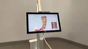
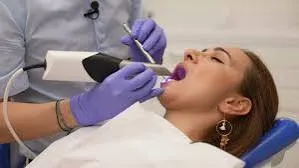
Intraoral scanners are advanced optical reading devices structured to map the teeth and surrounding tissues three-dimensionally, taking thousands of frames per second and combining them. These devices, which replace traditional dough-like impression materials, work on the principle of light reflection, projecting the intraoral topography onto the computer screen in a colored, high-resolution manner. During the scanning process, the tip of the device is moved over the tooth surfaces, and the data obtained is transferred to the software instantly, forming a three-dimensional model. Since this technology eliminates the need to hold a foreign substance in patients’ mouths, it aims to make the process quite comfortable for individuals with a gag reflex. In centers using current infrastructure such as Dentocare Clinic, the digital measurements obtained through intraoral scanners can be sent instantly to the prosthesis laboratory via email or cloud systems, without the need for a physical courier process. The instant feedback system offered by the scanners allows the physician to correct any missing or insufficient area seen in the impression simply by re-scanning that specific area, eliminating the need to retake the entire impression from scratch.
| Comparison Criterion | Traditional Impression Methods | Digital Impression (Intraoral Scanners) |
|---|---|---|
| Material Used | Dough-consistency materials such as silicone or alginate | Optical light, digital sensors, special software |
| Data Transfer and Communication | Sent to the laboratory via physical courier | Data transfer over the internet within seconds |
| Size and Storage | Plaster models need to be stored on physical shelves | Digital files are archived in cloud storage |
| Patient Comfort | Nausea and taste sensitivity may occur due to the impression material | Since there is no material contact, the gag reflex is not triggered |
| Error Detection and Correction | The entire procedure must be repeated if the impression is distorted | Only the missing area can be rescanned and added to the file |
In Which Processes Are CAD/CAM Systems Preferred?
CAD (Computer Aided Design) and CAM (Computer Aided Manufacturing) systems represent a technological integration that automates the modeling and casting processes done by hand in a laboratory setting. Data prepared digitally by the physician or transferred digitally to the laboratory is designed using CAD software in a way that is suited to the patient’s facial features, chewing forces, and the anatomy of neighboring teeth. Once the design is approved, thanks to the commands sent to the CAM unit (milling devices), zirconium, porcelain, or hybrid ceramic blocks are shaped by being carved with milling tips. Dentocare Clinic physicians make use of the data from these systems in the planning of patients applying in Kahramanmaraş, aiming to reduce the microscopic deviations that could arise from human hands. These technologies are frequently used in the production stages of crowns, bridges, porcelain veneers (laminate porcelain), and porcelain fillings called inlays/onlays. The wax modeling, investing, firing, and casting stages, which could take days in classic laboratory processes, become shapeable within hours thanks to CAD/CAM milling machines. Thus, the anatomical structure of the prostheses, their occlusion (bite) relationships, and margin fit can be calculated with computer parameters at much more predictable standards.
What Are the Functions of 3D Printers in Dentistry?
Three-dimensional printers are devices that, unlike CAM milling machines that work on a subtractive manufacturing logic, work on the principle of additive manufacturing, forming objects by hardening special liquid resins with light. In dentistry, they have a wide range of uses, especially in the production of models that directly reflect patients’ intraoral structure, the preparation of surgical guide templates, the printing of orthodontic clear aligners, and the production of temporary teeth. When digitally designed data is transferred to a 3D printer, the device forms the physical model by adding layers with a thickness of fractions of a millimeter on top of each other. When planning processes in orthodontics in which teeth are moved stage by stage, the model of each stage is produced from these printers and clear aligners are printed on top of them. In implant surgery planning, after the bone thickness is measured via tomography and the angle and depth of the implant are set digitally, the surgical guides that will direct the physician to this position during the operation are produced with 3D printers. This reduces the possibility of deviating from the planned angle, increasing the predictability of the surgical operation. With developing resin technologies, research continues not only on auxiliary materials but also on producing, with three-dimensional printers, some biocompatible restorative materials intended for permanent use.
What Dimension Does Digital Tomography (CBCT) Bring to the Diagnostic Process?
Cone Beam Computed Tomography (CBCT), unlike traditional two-dimensional panoramic X-rays, is an advanced imaging method that allows the patient’s jaw, tooth, sinus, and nerve canal structures to be examined three-dimensionally, in anatomical cross-sections. Since structures overlap in two-dimensional X-rays, clearly measuring certain anatomical details, infections, or bone thicknesses can become difficult. However, thanks to CBCT technology, the height, width, and density of the bone structure can be analyzed to the millimeter. Especially before implant surgery, the exact position of the nerve canal passing through the lower jaw (canalis mandibulae) or the extent of sagging of the sinus cavities in the upper jaw is determined with digital tomography, and the process is planned accordingly. Dentocare Clinic, which operates within the boundaries of Kahramanmaraş, makes use of this three-dimensional data in order to evaluate patients’ anatomical structures in detail and reduce surprise factors. The level of bone resorption in extracted tooth areas, the proximity of wisdom teeth to nerves, jaw joint (TMJ) problems, or the actual size of cysts at the root tip are determined by examination in cross-sections. This technology helps the physician virtually carry out the planned procedure on a computer screen, select material of the appropriate size, and detail the operation map.
How Is Smile Design Shaped in the Digital Environment?
Digital smile design is the process of evaluating characteristic features of the patient, such as facial features, lip structure, gumline, pupils, and skin tone, as a whole and adjusting asymmetries through computer software. This process is carried out not only with intraoral data but also blended with professional photographs of the patient taken from different angles and video recordings showing lip dynamics during speech. The intraoral digital scan data obtained and the studio photographs are superimposed on each other in a special design program. The physician completes the modeling on the photographs by selecting tooth shapes suited to the patient’s gender, age, and characteristics from a virtual library. The most distinctive feature of this application is that, before the procedure even begins, the final appearance of the teeth on the face can be presented to the patient on digital screens. In addition, after the design is complete, temporary plastic trial materials called a “mock-up” are placed on the patient’s teeth without any procedure being performed, allowing functional and aesthetic trials of the process to be done intraorally. In this way, the patient and physician reach a common ground within the framework of expectations and anatomical limits, clarifying the treatment roadmap.
| Reference Point | Effect on the Design Process |
|---|---|
| Interpupillary Line (the line between the pupils) | Determines the alignment and inclination of the teeth on the horizontal plane. |
| Midline (the vertical symmetry axis of the face) | Ensures that the meeting point of the two front incisors is centered on the face. |
| Lip Line and Smile Line | Balances the visible tooth length and amount of gum shown when smiling. |
| Zenith Points (the highest points of the gumline) | Leveling is planned to equalize the asymmetric appearance of the gums. |
| Functional Dynamics | The sounds produced during speech (F, V, S sounds) are tested against tooth lengths. |
What Innovations Have Digital Technologies Brought to Orthodontic Practice?
Orthodontic applications are a field focused on correcting dental crowding and bite problems. Processes that in past years proceeded using metal wires and brackets have today taken on a different dimension with digital planning software and clear aligner systems. When the three-dimensional measurements taken with an intraoral scanner are uploaded into orthodontic software, all the movements of the teeth from their starting position to their final position are calculated to the millimeter. Through animations created in the computer environment, this process, spread over months, can be played back virtually, and the outcome can be evaluated in advance. In line with these calculations, successive series of clear aligners that will apply force to bring the teeth to the determined position are produced. In centers that integrate modern technological developments into their practice, such as Dentocare Clinic, these clear aligner technologies, which help orthodontic patients complete the process without experiencing physical difficulties such as wire or bracket irritation, are frequently considered. Thanks to digital software, not only the movements of the teeth but also the effects of these movements on the roots and jawbone are analyzed, supporting more stable planning. Since patients can remove their aligners while eating or brushing their teeth, they can maintain their oral hygiene habits in a standard manner.
Why Are Digital Shade-Matching Devices (Spectrophotometers) Used?
In dental restorations, it is of great aesthetic importance for the porcelain or zirconium material to be made to match the natural tooth color exactly. Traditionally, physicians determine the color by holding the shade guides provided by laboratories next to the patient’s tooth and making use of ambient light, judging by eye. However, factors such as the lighting of the environment, different times of day, reflection of wall colors, or eye fatigue can lead to differences in color perception. Digital color measurement devices (spectrophotometers) are used to eliminate these variables. When the device is brought into contact with the tooth surface, it analyzes the light reflected from the tooth’s enamel and dentin layers using its own standard LED light source. It turns not just a single color of the tooth, but the color transitions in the cervical, middle, and incisal areas, the degree of translucency, and brightness values into mathematical data. This map obtained is sent digitally to the laboratory technician, making it easier for the material to be produced to reasonably match the natural tooth’s light transmission and color depth.
How Does the Digital Dentistry Workflow Affect Laboratory Communication?
The communication network between the clinic and the prosthesis laboratory has a direct effect on the quality of the resulting material. In the classic workflow, the process of sending impressions by courier, pouring them into plaster at the laboratory, preparing the model, and having it delivered back to the clinic via courier is a cycle that can take days. During this period, dimensional changes in the impression material or breakage in the plaster models during transport can occur. In the digital workflow, the process instead proceeds through cloud-based systems. The moment the physician scans the patient, the data lands on the laboratory’s screen within seconds. By opening the design software, the technician can carry out analyses on the model simultaneously with the physician, or over the phone, evaluate the margin lines (tooth cutting lines), and, if there is any issue, ask the physician to rescan before the patient has even left the chair. The digitalization vision of establishments such as Dentocare Clinic, which serves the Kahramanmaraş area, includes adopting these communication protocols that minimize time loss and allow for instant intervention. At the same time, since patients’ past data can be stored undamaged in digital archives, it becomes possible to produce a new item in the same shape based on the old files in the event of a fracture or a need for prosthesis replacement in later years.
Frequently Asked Questions (FAQ)
We aim to comprehensively answer the questions that arise in patients’ minds about the use, processes, durations, and materials of digital technologies in the field of oral and dental health. Below, you can find the main topics of curiosity regarding digital dentistry applications in a question-and-answer format.
1. How long do digital dentistry applications take?
The duration of digital dentistry processes varies depending on the nature of the procedure to be applied, the number of materials to be produced, and the in-house production (milling) capacity available at the clinic. Impression, laboratory stage, and fitting processes, which could take days or weeks in traditional methods, can be reduced to a single session with the integrated use of CAD/CAM systems and intraoral scanners. If the clinic has production units, the time from data acquisition to design and the material being milled and applied to the patient can be completed within a few hours. However, in multiple tooth restorations or in laboratory production processes originating abroad, such as clear aligners, this period may extend depending on the planning.
2. Does nausea occur when taking a digital impression?
One of the main goals in the development of digital impression systems is to increase the level of comfort for patients during impression-taking. While the large impression trays placed in the mouth in the traditional method and dough-consistency materials that can tend to flow toward the throat can stimulate the gag reflex, digital impression devices (intraoral scanners) work with a small camera tip. These optical readers, which are moved only over the tooth surfaces without using any material, do not press on the palate or throat area, helping individuals with a gag reflex get through the process comfortably.
3. Are the crowns made with CAD/CAM technology durable?
Restorations prepared with computer-aided manufacturing (CAM) systems are generally milled as a single piece (monolithic) from industrially compressed, homogeneous zirconium, lithium disilicate, or hybrid porcelain blocks under high pressure. Compared to layered porcelain that is hand-shaped and fired in a laboratory, these blocks structurally carry less risk of micro-cracks or air bubbles. The physical strength offered due to the nature of the material is designed with long-term use against chewing forces in mind. In addition to material quality, the design and tooth-preparation principles applied by the physician positively affect durability.
4. In which areas are 3D printers used in dentistry?
Three-dimensional (3D) printers are actively used in quite a wide range of areas in dentistry. They are used in printing the models from which successive clear aligners that move the teeth will be produced in orthodontic treatments, in preparing surgical guides that support the planned execution of implant operations, and in the quick production of temporary teeth to be applied to the patient. They also offer functional solutions in areas such as the production of night guards, the printing of mock-up models for smile design trials, and the preparation of jaw surgery training models.
5. Can digital smile design be applied to every patient?
Digital smile design is a planning-focused concept that can be applied to many adult individuals whose oral and dental development is complete. People who have shape, size, or color asymmetry in their teeth, mild crowding, or irregular gum levels can benefit from this analysis. Since the patient’s facial structure is used as a reference during the design process, the resulting outcome is shaped individually for the person. However, in individuals with serious skeletal problems in the jawbones or an advanced degree of orthodontic disorder, it may be considered necessary to carry out infrastructural adjustments, such as surgery or wire treatment, before an aesthetic restoration.
6. What is the radiation level of digital X-rays?
Thanks to developing sensor technologies, the radiation dosage used in digital X-ray and tomography (CBCT) devices has been greatly limited compared to old-type analog (film) X-ray devices. Since digital sensors are much more sensitive to X-rays, the exposure time needed to form the image is quite short. Cone beam 3D tomography devices also target a much narrower area compared to medical tomography devices, sending the beam in a cone shape, thereby minimizing the dosage received by the patient. Since the quality of the data obtained provides very important details for diagnosis compared to the dose given, it is frequently preferred in accordance with radiology standards.
7. How can digital dentistry services be accessed in Kahramanmaraş?
It is possible to access these services in the Kahramanmaraş area through clinics that support their technological infrastructure with current systems. Dentocare Clinic responds to this need by housing three-dimensional planning tools such as CAD/CAM systems, intraoral optical scanners, and digital tomography within its facilities for patients’ diagnostic and treatment processes. When patients apply to the clinic, they are informed in detail about processes planned through the digital workflow instead of conventional methods, and can see transparently how their own treatment roadmap will be created in the digital environment.
8. Can intraoral scanners be used in children?
The use of intraoral scanners is not limited by age and helps processes go much more comfortably, especially in child patients (pediatric dentistry). Children’s gag reflexes or fear of impression material can make their cooperation in the dental chair more difficult. Since the scanner’s digital camera tip stays in the mouth for a short time and produces colorful, animated images, it captures children’s interest and helps them perceive it as a kind of game. This makes it more practical to collect fast and detailed data from children who have difficulty waiting still.
9. What is the process for zirconium crowns with digital methods?
When the zirconium crown process is done with a digital workflow, it begins with the dentist performing the necessary reduction (preparation) procedure on the relevant tooth, followed by taking an optical impression with an intraoral scanner. This digital impression obtained is transferred to CAD software, and the shape of the tooth, its chewing surfaces, and its relationship with the neighboring teeth are designed in a computer environment. The design data is transmitted to the CAM milling device, and the zirconium blocks are shaped through the milling process. The milled zirconium goes through a sintering (firing) stage in special ovens to increase its durability, and is colored to be made compatible with the patient’s mouth.
10. How does digital dentistry contribute to implant planning?
The heart of implant surgery is the accurate evaluation of the anatomical structure of the jawbone. Digital dentistry performs millimeter-precise analysis of the jawbone with three-dimensional tomography (CBCT). The physician measures bone density in a virtual environment, determines the appropriate implant diameter and length, and safely calculates the distance to nerve tissue and sinus cavities. In line with this data, surgical guides prepared with a 3D printer guide the physician during the operation, allowing the implant to enter only at the planned angle and depth. In this way, the process is planned in a way that is free of surprises, predictable, and in accordance with standards.
11. What is the role of digital technologies in the production of night guards?
Night guards, which are focused on protecting tooth surfaces against bruxism (teeth clenching and grinding), can be produced much more ergonomically and precisely with digital systems. Based on the digital impression taken with intraoral scanners, the patient’s bite disorders are examined in CAD software, and the thickness of the guard and its contact points with the teeth are adjusted to the millimeter. Night guards produced afterward with 3D printers or milling units from special biocompatible clear materials retain their shape longer compared to traditional vacuum-formed soft guards and carry the hardness characteristics needed to support the jaw joint (TMJ).
12. What materials are used in digital dentistry?
Digital manufacturing systems develop in parallel with materials science. In CAM systems (milling machines), zirconium oxide blocks, lithium disilicate (E-max, etc.) glass ceramics, resin nano ceramics, PMMA (polymethyl methacrylate) temporary materials, and hybrid composite blocks are frequently used. On the 3D printer side, surgical guide resins, castable resins, model resins, temporary crown materials, and splint/night guard production liquids are preferred. The common feature of these materials is that, thanks to their homogeneous structure, they can offer industrial strength to factory-produced standards.
13. Is it possible to see the result in the digital environment before treatment?
Yes, one of the biggest advantages of digital planning is simulation and mock-up applications. In veneer, zirconium, or orthodontic clear aligner treatments where aesthetic expectations are high, computer software presents the final result by modeling it three-dimensionally on screen based on the initial data obtained. The patient can see on the screen how their teeth will be arranged and what their shape will be after treatment. In applications such as smile design, this digital data can also be turned into a physical model with a 3D printer and attached to the patient’s teeth with a temporary material, allowing a realistic trial to be done in front of a mirror.
14. How are digital technologies used in orthodontic processes?
Orthodontic diagnosis and data analysis have taken a leap forward thanks to digital software. Traditional cephalometric (lateral skull) X-ray tracings and angle measurements are calculated automatically by digital programs, determining bone growth direction and dental deviations. Based on these analyses, integrated with intraoral scan data, if wire treatment is to be done, custom-made bracket positions can be planned virtually and bonded to the tooth all at once using transfer trays. These methods optimize the physician’s chairside working time while increasing the predictability of the treatment mechanics.
15. How is digital planning done for clear aligner treatments?
Clear aligner treatment (wireless orthodontics) is a system built entirely on a digital workflow. After the patient’s intraoral optical scan is done, this data is uploaded to the cloud. With the support of special software, artificial intelligence, and the physician’s direction, the teeth, along with their roots, are gradually moved from their current position toward the ideal alignment they should be in. If the process consists of, for example, 20 stages, the computer creates a separate three-dimensional model for each stage, and 20 clear aligners suited to these models are produced. These aligners, which the patient changes on a weekly basis, manage the process by transferring the micro-movements planned by the software to the teeth in sequence.