What Is Digital Dentistry?
Digital dentistry is a dental approach in which intraoral measurements, diagnostic stages, and prosthesis production processes are carried out through computer-aided technologies, three-dimensional scanners, and medical software. This system transfers data to a digital environment using optical cameras instead of standard measurement materials, allowing the planning process to be managed through computer screens.
While traditional records are based on physical measurement materials being held in the mouth for a certain period, in current systems these stages are completed in a short time using optical readers. The data obtained is sent as a data packet directly to the laboratory environment or to in-clinic production devices (milling units). This helps minimize the possibility of dimensional changes that could arise from the physical properties of measurement materials.
The main components of the system consist of the following:
- Intraoral Scanners: Cameras that record the three-dimensional topography of the teeth and gums.
- Design Software (CAD): Programs in which the data coming from the scanner is processed and the shape of the prosthesis is virtually drawn.
- Manufacturing Devices (CAM): Units in which the crown, bridge, or plate whose design has been completed is turned into a physical object, either by being milled from porcelain/zirconium blocks or printed with 3D printers.
In Which Areas Is Digital Dentistry Used?
This system is used in different disciplines, such as crown and bridge fabrication in prosthetic dental treatments, orthodontic planning carried out with clear aligners, the preparation of surgical guide plates, and aesthetic planning. With the expansion of the computer-aided manufacturing network, medical software can be integrated into many diagnostic and restoration processes within the clinic.
The data-processing capacity of the applied technology not only records the current condition but also allows the treatment outcome to be simulated virtually in advance. How a given procedure will affect the bite of the teeth or the facial anatomy can be calculated on medical screens before treatment begins.
| Area of Use | Function of the Digital System | Output Obtained |
|---|---|---|
| Prosthetic Treatments | Virtual design of porcelain, zirconium, or full ceramic crowns/bridges. | Dental prostheses milled from blocks with millimeter-precise fit. |
| Orthodontics (Clear Aligners) | Simulation of tooth movements in weekly periods via software. | Series of 3D-printed clear aligners that gradually guide the teeth. |
| Surgical Applications | Determining the position of the titanium root by combining bone volume with tomography data. | Medical guide plates that direct the surgical drills to the planned angle. |
| Smile Design | Proportional shape planning by matching facial photographs with intraoral scans. | Virtual models shaped to fit the individual’s anatomy. |
How Does Digital Impression-Taking Differ from Traditional Methods?
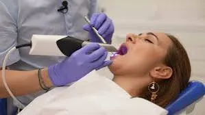
In the traditional method, dough-consistency impression materials placed in the mouth are replaced, in digital impression-taking, by optical cameras that capture thousands of photographs within seconds. These optical scanners eliminate the need for physical impression materials by instantly transferring a three-dimensional model of the teeth and surrounding tissues to the computer screen.
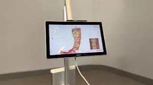
The preparation of physical impression materials, the holding of trays placed in the mouth for a period, and the setting phase can stimulate the gag (nausea) reflex in individuals. Optical scanners, on the other hand, work by being moved over the tooth surfaces with the help of a small-sized tip. During this process, the colored, three-dimensional topography of the teeth begins to form instantly on the computer screen.
The process of delivering the measurements to the laboratory environment also differs. While traditional impressions are transported via couriers, optical records are uploaded to medical systems over the internet network. The laboratory technician can open the model on their computer immediately after the impression is taken and move on to the design stage.
How Does CAD/CAM Technology Contribute to the Workflow Process?
CAD (Computer-Aided Design) and CAM (Computer-Aided Manufacturing) systems enable the digital impressions taken to be shaped three-dimensionally and then produced from porcelain or zirconium blocks using milling devices. The physician draws the shape of the restoration on the computer, and the device turns this design into reality by machining a physical block.
CAD software detects the anatomical boundaries of the tooth and suggests to the physician a shape in which chewing forces will be distributed evenly. Based on this suggestion, the physician refines the contact points of the tooth with neighboring teeth, its bite relationship with the opposing jaw, and the gumline using digital tools. Once the design is complete, the data is transferred to the CAM unit.
CAM units are devices equipped with diamond or carbide tips called mills. Raw zirconium, lithium disilicate, or glass ceramic blocks placed into the system are carved three-dimensionally according to the code coming from the software. As a result of this carving process, the designed crown or filling shape is physically revealed.
How Are Digital Applications Planned at Kahramanmaraş Dentocare Clinic?
Dentocare Clinic, which serves the Kahramanmaraş area, carries out the steps from the diagnostic stage to prosthesis production using intraoral scanners and three-dimensional tomography systems. The individual’s anatomical data is transferred to digital platforms, so that the shape and dimensions of the procedures to be performed are assessed on a computer by both physician and patient before the procedure.
For individuals applying to the clinic, the prosthetic planning process begins with the scanning of the lower and upper jaws using optical cameras. The bite registration (the position in which the teeth touch each other) is likewise taken digitally. The tomography data and intraoral scan data of patients in the Kahramanmaraş area are brought together in medical software by Dentocare Clinic physicians. This integration allows the thickness of the jawbone and the gum shape on top of it to be evaluated simultaneously during surgical procedures.
The production of the planned restorations is directed to the appropriate devices depending on the type of material and the scope of the procedure. Recording the process with medical software creates a structural standard whereby, in the event a prosthesis replacement is needed in the future, the old records can be recalled from the archive to reproduce the same shape.
For What Purposes Are Three-Dimensional (3D) Printers Preferred in Dentistry?
Three-dimensional dental printers are preferred for turning optical impressions into physical models, printing guide plates for surgical interventions, producing temporary tooth crowns, and preparing base models for series of clear orthodontic aligners. Unlike the milling method, these devices produce output by hardening liquid resins layer by layer.
While milling units work by removing material from hard blocks, 3D printers work on the principle of additive manufacturing. Resin-based liquid material is cured with light sources, forming layers with a thickness of a very small fraction of a millimeter. When each layer stacks on top of the others, the final product emerges.
| Material Produced | Technology Used | Purpose of Use |
|---|---|---|
| Dental Model | Resin-Based 3D Printer | Being able to carry out laboratory work on a physical copy of the patient’s mouth structure. |
| Surgical Guide | Biocompatible Clear Resin | Supporting the placement of titanium artificial roots into the bone at the targeted angle and depth. |
| Temporary Crown | Tooth-Colored Resin | Ensuring the patient does not go without teeth and that chewing function continues during the production process of the final prostheses. |
How Does the Digital Smile Design Process Proceed?
The digital smile design process proceeds by recording the individual’s facial photographs, lip structure, and speech dynamics, and combining this data with intraoral scan models in medical software. In line with these reference points, a tooth shape suited to the facial features is designed virtually and presented to the patient.
When planning a smile, anatomical reference points such as the horizontal line passing through the pupils, the nostrils, lip symmetry, and the midline of the face are taken as a basis. The software places new virtual tooth shapes over the existing teeth. The finished result of the planned porcelain veneers, zirconium crowns, or gum-leveling procedures is visualized on screen without any intervention being performed on the teeth.
Once the virtual design is complete, it is turned into a physical model using three-dimensional printers. Using special molds (the mock-up technique), this resulting shape is copied onto the patient’s mouth with a temporary material. The individual has the opportunity to evaluate the planned tooth structure on their own face in the mirror, and if a change to the shape is requested, revisions are made by returning to the software.
What Are the Frequently Asked Questions?
1. Does the gag reflex occur during digital impression-taking?
The large trays and dough-consistency impression materials used in standard methods can stimulate the gag reflex when they touch the palate. Optical scanners, on the other hand, reduce the rate at which this reflex occurs, since they do not involve any dough in the mouth and work with a small tip.
2. Does computer-aided manufacturing apply only to zirconium?
CAD/CAM systems are used not only for zirconium but also for producing lithium disilicate (e.max), full ceramic (porcelain), resin nano ceramic, composite blocks, and metal substructures. The block to be used is selected according to the physician’s diagnosis.
3. Do the scanner cameras emit radiation?
Intraoral scanner cameras are not radiographic (X-ray) devices. Since their working principle is based on light reflection and optical reading, similar to a photo camera or camcorder, there is no radiation emission involved.
4. How is digital planning included in the orthodontic process?
Optical records taken during orthodontic treatment transfer the position of the teeth to the software. The physician plans, in a digital environment, the steps needed for the movement of the teeth. Planning is carried out by producing clear aligners corresponding to each of these steps.
5. Which materials can be produced with three-dimensional printers?
Using dental 3D printers, it is possible to produce temporary crowns, surgical guide templates, base models used in the production of clear aligners, splints (night guards), and impression trays.
6. How is color selection made in the digital environment?
Modern intraoral scanners have special lenses and spectrophotometer technology that can analyze the actual color of the tooth while also recording its topography. This data is also confirmed with standard shade guides before being sent to the laboratory.
7. How long does the intraoral scanning procedure take on average?
Although it varies depending on the condition of the case, saliva flow, and the position of the teeth, a full scan of a single jaw is generally completed within 1 to 3 minutes under routine clinical conditions.
8. Is every case suitable for digital production?
The great majority of cases can be scanned with this system. However, in cases with heavy bleeding of the gum tissue or in deep areas that the scanner has difficulty reaching, support with traditional methods can be provided at the physician’s discretion.
9. How are surgical guide plates prepared in the digital system?
The patient’s three-dimensional bone tomography (CBCT) and intraoral optical scan data are superimposed in the software. The structure of the bone and the shape of the gum are evaluated simultaneously, the position of the artificial root is drawn, and a clear guide plate that will direct this position is designed.
10. Can temporary crowns be produced with CAD/CAM systems?
After the impression is taken, in order to prevent the patient from going without teeth until the permanent restoration is produced, anatomical temporary teeth can be produced either by milling from PMMA blocks or with resin-based 3D printers.
11. Can traditional impressions be transferred to the digital environment?
In cases where impressions are taken with physical dough, the resulting plaster models can be scanned using desktop scanners found in laboratories and transferred into the CAD/CAM system.
12. How are the gum margins determined with optical scanners?
As in the traditional method, medical cords are placed between the tooth and the gum to push the gum away slightly. The camera scans this gap and records the finishing margin of the prosthesis in the software, aiming for the compatibility of the structure to be produced with the tissue.
13. What is the purpose of face scan technology?
This technology, which transfers the three-dimensional anatomy of the individual’s entire face to the computer, helps with aesthetic planning in extensive front-region restorations by taking as reference the compatibility of the tooth shape with lip support, the shape of the nose, and eye alignment.
14. How does the system record the bite (occlusion)?
The physician asks the patient to bite their teeth in their normal position, and scans, with the scanner camera moved along the side cheek areas, the parts where the teeth make contact with each other. The software automatically superimposes the lower and upper jaw scans based on this bite record.
15. How does the physician transmit the digital design to the laboratory?
The scan data and photographs are saved as files with special extensions. These files are sent to the relevant prosthesis production laboratory through encrypted systems or cloud software that complies with patient confidentiality.