Why is the Jawbone Important in Dental Implant Applications?
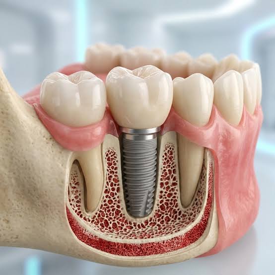
In implant applications planned to replace missing teeth, the jawbone is at the center of the process because it serves as the main foundation where the placed artificial root can anchor. Just as the stability of a building depends on the quality of the ground on which it is built, an implant’s ability to perform its functions inside the mouth depends on the volume, density, and general health condition of the surrounding bone tissue. Our natural teeth are anchored in their special sockets within the jawbone via their roots, directly transmitting the forces generated during chewing to the bone; this mechanical transmission encourages the bone tissue to remain alive and preserve its form. When a tooth is lost for any reason, the bone tissue in that area tends to undergo a volumetric decrease over time as it no longer receives chewing stimuli. At this point, the implant acts as a biocompatible root that not only fills a visual gap, but also sends mechanical stimuli to the jawbone, supporting the slowing down of bone resorption. For an ideal healing process to occur and for the titanium material to biologically integrate with human tissue (osseointegration), healthy bone tissue of sufficient height and width is required in that region. Every step of the process requires a detailed analysis of the individual’s anatomical structure and the creation of a roadmap tailored to their bone capacity.
What is the Role of the Jawbone in Implant Treatment?
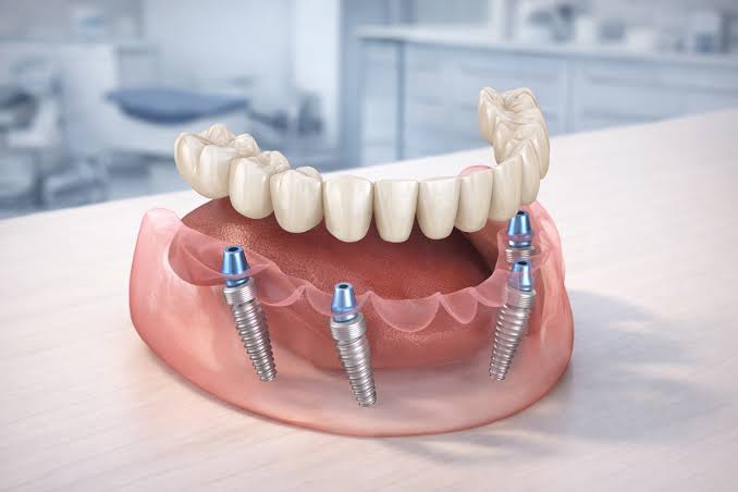
The role of the jawbone in implant treatment is to physically surround the inserted titanium screw, provide it with stability, and evenly distribute the pressure generated during chewing to other areas of the mouth. Titanium is a special material that is not perceived as a foreign body by the human organism, allowing bone cells to adhere to it and proliferate. During the recovery period of several months following the procedure, bone cells settle into the micro-roughened surfaces of the implant, forming a virtually biological weld. The desired level of this integration depends directly on the structural quality and volume of the bone. If the bone tissue is weak, thin, or shallower than necessary, the placed artificial root may not demonstrate sufficient resistance against chewing forces. In the long term, this can cause sensitivity in the tissues surrounding the implant or lead to the structure losing its stability. Therefore, the jawbone is a vital foundation that plays a continuously supporting role not only at the beginning of the process, but also to ensure that the application functions in the mouth for many years.
What Changes Occur in the Jawbone After Tooth Loss?
The most noticeble change in the jawbone following tooth loss is that the bone tissue in that area gradually begins to lose volume due to its loss of function. Bone tissues in the human body work on the “use it or lose it” principle; just as an immobile muscle weakens over time, a jawbone that receives no chewing pressure starts to thin and resorb. During the first six months following tooth extraction, a significant reduction in both horizontal and vertical directions is observed in the bone surrounding the extraction socket. Although this narrowing slows down in subsequent years, it can reach an extent that affects the balance of the entire dental arch as adjacent teeth lose support. Additionally, the nerve canals in the lower jaw and the sinus cavities in the upper jaw draw closer to the gums as bone volume decreases, narrowing the anatomical distances. Since these physical changes can complicate future replacement procedures, it is recommended to take steps to restore function without letting too much time pass after tooth loss.
| Duration of Tooth Loss | Observed Condition in the Jawbone | Clinical Implication |
|---|---|---|
| First 3 – 6 Months | A rapid horizontal (width) bone reduction process begins. | The extraction site becomes sunken, and a slight inward collapse of the cheek and lip tissues may be observed. |
| 1 – 3 Years | Vertical (height) bone loss begins to become pronounced. | Adjacent teeth tend to tilt toward the gap. |
| 3 Years and Above | Significant reduction in bone volume and convergence of anatomical structures. | Additional supportive procedures such as bone grafting (bone powder) are required prior to implant application. |
Under What Circumstances Does Jawbone Deficiency Occur?
Jawbone deficiency occurs as a result of various factors such as prolonged edentulism (toothlessness), advanced periodontal disease (periodontitis), cyst or tumor surgeries, difficult tooth extractions, and long-term use of ill-fitting removable dentures. When gum inflammation starting in the mouth deepens over time and reaches the bone tissue supporting the tooth, it can disrupt the structural form of the bone in that area and lead to its resorption. If a tooth is lost due to this reason, the remaining foundation is already weakened. Similarly, in individuals who have worn full dentures for many years, the constant pressure of the denture on the gums and underlying bone affects tissue nutrition, causing a decrease in bone height. Difficult tooth extractions affecting surrounding tissues can also damage the bone walls in that area, leading to volume loss during the healing process. Having an anatomically thin jaw structure can also bring about a deficiency state as a hereditary factor.
How Do You Know If There Is Sufficient Bone for an Implant?
Whether there is sufficient bone for an implant is understood by conducting a detailed analysis using advanced radiological imaging techniques, such as 3D cone-beam computed tomography (CBCT), alongside an intraoral clinical examination. While standard two-dimensional panoramic X-rays give an idea only about the height and general condition of the bone, 3D tomographies allow millimetric measurements of the bone’s width, density, the distance to the nerve canal in the lower jaw, and the location of the sinus cavities in the upper jaw. These digital analyses provide critical data for determining the diameter and length of the artificial root to be placed. In the evaluation processes carried out within Dentocare, individuals’ unique anatomical structures are examined through these digital plans to create a roadmap based on the resistance the bone will exert. Upon reviewing the imaging results, the dentist can determine if the bone is adequate, or, in case of deficiency, plan additional steps to reinforce the area within the same session.
In Which Cases is the Bone Graft (Bone Powder) Procedure Preferred?
The bone graft (bone powder) procedure is preferred when there is insufficient height or width in the jawbone to support an implant, aiming to increase the volume of that region by supporting it with biological materials. The deficient area is filled sometimes using a piece of bone harvested from the patient’s own body, and sometimes using sterile, biocompatible graft materials prepared in a laboratory environment. During the procedure, the bone graft is placed in the deficient area, and the area is covered with a special resorbable barrier (membrane). Over the months, the body uses this material as a scaffold, transporting its own living bone cells to the region and transforming the artificial material into real bone tissue. This process is an important preparatory step taken to strengthen the weakened foundation and ensure that the placed artificial root functions in a durable environment for years to come.
Major Factors Creating the Need for a Bone Graft:
- Many years having passed since tooth extraction and the thinning of bone walls.
- Damage caused to the jawbone by advanced periodontal diseases.
- Bone defects formed during tooth extraction or gaps remaining after cyst removal.
- Sinus cavities drooping in the upper jaw posterior region, reducing bone height.
Why is a Sinus Lift Operation Performed?
A sinus lift operation is performed when, following the loss of molars in the posterior region of the upper jaw, the air cavities positioned directly above the teeth (maxillary sinuses) sag downward, reducing the bone height required for placing an implant. Sinuses are natural air chambers with functions such as respiration and voice resonance. While teeth are present, these chambers are supported by bone tissue; however, after teeth are extracted and bone volume decreases, the sinus membrane gradually expands downward. To prevent the artificial root from protruding into this cavity and damaging the sinus membrane, the membrane is gently pushed upward with special instruments, and bone powder is placed in the created space. Thanks to this operation, adequate vertical bone height is recreated in the posterior upper jaw, preparing a healthy foundation for implant placement.
Can Implants Be Applied to Individuals with Osteoporosis?
In individuals with osteoporosis (bone loss), implant applications can be performed provided that approval is obtained from the physician attending the patient and necessary additional precautions are taken; however, the process is planned more meticulously, considering the metabolic rate and healing potential of the bone. Osteoporosis is a systemic condition that leads to a reduction in bone density throughout the entire body. The outer hard layer (cortical bone) in the jawbone of these patients may be thinner than normal, and the inner spongy layer may have larger spaces. Prior to the procedure, dentists analyze the bone quality and select materials that possess an appropriate surface structure with high anchorage capacity even in weak bone. Since certain specific drugs used in osteoporosis treatment (bisphosphonate group) can affect bone healing, obtaining a detailed medical history of the patient is of great importance for managing the process.
How Do Systemic Diseases Affect Jawbone Quality?
Systemic conditions such as diabetes, thyroid issues, or autoimmune disorders directly affect general blood circulation and cellular healing speed, thereby playing a decisive role in jawbone quality and its resistance against infections. Uncontrolled diabetes, in particular, reduces the amount of oxygen reaching tissues, slowing down the nutrition of bone cells. This situation can cause the biological integration process between the placed artificial root and the bone to take longer than expected. However, these conditions do not pose an obstacle to treatment. When the individual maintains their health values at normal levels under the supervision of relevant medical specialists, the healing potential of oral tissues largely recovers. The key is to establish a gradual and controlled treatment schedule tailored to the patient’s current medical status through transparent information sharing between doctor and patient.
How Do Bone Evaluation Processes Progress at Dentocare Clinic?
Bone evaluation processes at Dentocare Clinic progress by examining the individual’s oral and jaw structure with the aid of 3D digital technologies to comprehensively map current volume, density, and overall oral health status. After listening to the patient’s complaints and expectations, data obtained through advanced tomographic analyses are transferred to a digital platform. At this stage, the angles and dimensions of the roots planned for placement are simulated on a computer. This meticulous preparation phase helps prevent unexpected situations during surgery while aiming to establish a sustainable chewing function compatible with the patient’s anatomy. In cases where bone tissue is weak, Dentocare dentists inform the patient, sharing the necessity of preparatory procedures such as bone grafting or sinus lifting in detail, and create a personalized timeline.
What Should the Diet Be Like During the Bone Healing Process?
During the bone healing process, the diet should revolve around a menu rich in protein, calcium, and vitamins to supply the building blocks required by tissues, while care should be taken to prefer soft foods that will not mechanically stress the surgical area. For the first few days following the procedure, it is recommended to stay away from hard and crunchy foods that can strain the jaw muscles. Yogurt, warm soups, well-boiled vegetables, and pureed foods support the protection of the blood clot structure in that area. Consuming foods rich in vitamin C and vitamin D enables the body to perform bone synthesis, serving as a supporting factor for healing potential. Additionally, ensuring foods are neither too hot nor too cold helps reduce tissue sensitivity, and increasing water intake is recommended to prevent food particles from lodging in suture sites.
How Can Jawbone Loss Be Prevented After Implant Placement?
Jawbone loss after implant placement can be kept under control by paying meticulous attention to oral hygiene, not neglecting interdental cleaning, and adhering to routine dental check-ups at specified intervals. The development of gum infection around the artificial root (peri-implantitis) is one of the most fundamental risk factors leading to bone loss. Just like with natural teeth, accumulated bacterial plaque from inadequate brushing can progress from the gums to the bone, causing resorption in surrounding tissues. To prevent this, using soft-bristled toothbrushes and thoroughly cleaning the area with interdental brushes or specialized dental floss is of paramount importance. Avoiding habits that require excessive force (such as nighttime teeth grinding) or using a protective night guard for this condition reduces unbalanced loads on the bone, allowing bone health to be preserved for many years.
Is Jawbone Density Sufficient for Implants in Advanced Ages?
Although jawbone density in advanced age generally shows a slight decrease associated with the aging process, the status of bone quality varies depending not only on chronological age, but also on overall health, nutritional habits, and a history of long-term edentulism. Aging alone does not pose an obstacle. Many older individuals can benefit from the procedure smoothly because they possess a healthy metabolism and adequate bone quality. In cases where resorption has occurred over time, tissue can be augmented through bone grafting procedures offered by modern dentistry to create a suitable foundation for treatment. The age-related slowdown in bone tissue healing rate may only require the dentist to schedule a slightly longer waiting period; however, attaining a functional dental arch is possible across all age groups.
How Does Smoking Affect Jawbone and Implant Integration?
Smoking constricts blood vessels in the mouth due to its toxic substances and high heat, reducing the amount of oxygen and nutrients that need to reach the surgical tissue; this directly adversely affects the cellular integration process between the bone and the artificial root. Intensive blood flow is required for healthy healing. Impaired tissue vascularization due to smoking increases the risk of infection in the area while suppressing the body’s self-repair capacity. For this reason, it is recommended to limit or pause smoking before the procedure and during the several-month healing period. Even after the recovery period has passed, long-term smoking has the potential to weaken tissue defense mechanisms and lead to loss of bone support around the artificial root.
When is an Implant Placed After a Bone Graft?
The timing of implant placement after a bone graft varies depending on the volume of bone graft added to the region, the type of material used, and the rate at which the individual’s bone cells biologically integrate this material, but generally involves a waiting period of three to six months. If the bone deficiency is very minor, graft placement and insertion of the artificial root into its socket can be performed simultaneously in the same session. However, if a major volume augmentation or detailed site preparation such as a sinus lift has been performed, a maturation phase lasting several months is required for the added material to transform into solid, stable bone tissue. During this period, tissues are checked with regular X-rays; after confirming that bone density has reached the target level, the titanium root placement stage is initiated.
Frequently Asked Questions
1. Can I tell by myself if there is resorption in my jawbone?
Although visible signs such as receding gums or inward collapse of the lips in the missing tooth area may give an idea, actual bone volume can only be understood through evaluation by a specialist dentist using panoramic or 3D X-rays.
2. Is the use of bone powder (graft) a difficult process?
It is a preparatory procedure performed under local anesthesia. Because tissues are numbed during the procedure, it is comfortable; any mild swelling or sensitivity that may develop afterward can easily be controlled with ice compresses and supportive medications recommended by the dentist.
3. Does a sinus lift operation change my face shape?
No, sinus cavities are structural chambers located inside the upper jawbone. The intervention purely involves manipulating the membrane inside and does not cause any changes to facial contours.
4. Can an implant be placed immediately after tooth extraction?
If the bone walls at the extraction site are intact and there is no active infection, placement can be planned in the same session immediately after extracting the tooth. This situation is clarified following clinical evaluation.
5. What should I do if I am taking osteoporosis medications?
You need to inform your dentist in detail about the names and duration of use of the osteoporosis medications you are taking (especially bisphosphonates). Considering your medical history, the dentist will plan the process in coordination with your treating physician.
6. What should I pay attention to while the bone is integrating after the procedure?
Avoiding chewing hard foods that could exert pressure on the area, pausing smoking, and maintaining meticulous oral hygiene using interdental brushes are the key elements supporting the integration process.
7. How long does bone integration take?
A waiting period of approximately 2 to 3 months for the lower jaw and 3 to 4 months for the upper jaw (due to anatomical structure) is generally anticipated. If additional bone powder is used, this period may be extended by a few more months.
8. Is there a possibility of bone graft rejection by the body?
Medically prepared graft materials are highly biocompatible products sterilized with advanced technology. Since they integrate with the patient’s own cells to act as a structural scaffold, an allergic reaction or rejection is not an expected outcome.
9. Is my bone too weak because of advanced age?
Although advanced age causes bone to lose some density, weakness to a degree that would prevent the application is rarely observed. When necessary, supportive materials can be used to conduct the process smoothly.
10. Should I consume milk and dairy products after the procedure?
Calcium-rich foods contribute to tissue healing; however, during the first few days, soft purees at room temperature (not excessively hot), warm soups, and yogurt are ideal choices to protect the blood clot.
11. Can short implants be used if my bone is very thin?
Yes, short titanium screws with increased surface area, specially designed for cases with limited bone level, are available today. Your dentist will decide on this option through 3D measurements.
12. How do gum diseases cause bone resorption?
An infection starting in the gums, if left uncleaned, deepens to affect the connective tissue around the roots and subsequently the bone. The inflammatory environment impairs bone cell function, initiating resorption and tissue loss in that area.
13. Does titanium harm the bone?
Titanium is used as a prosthetic material in many fields of medicine due to its tissue-friendly (biocompatible) nature. The body does not perceive it as a foreign substance, and bone cells settle into the pores on its surface, establishing a natural bond.
14. I have used removable dentures for many years; has my bone resorbed?
Since removable dentures transmit chewing loads directly to the gums and underlying bone, volume reduction over time in that region is likely. This condition can be evaluated through clinical and radiological tests, and support can be provided to necessary areas.
15. Should the bone grafted area be brushed?
During the initial days, brushing is not performed to avoid contact with sutures in the surgical site; instead, chemical cleaning is provided using mouthwashes prescribed by the dentist. As healing progresses, you can return to brushing with gentle movements.