Fundamentals of Orthognathic Surgery
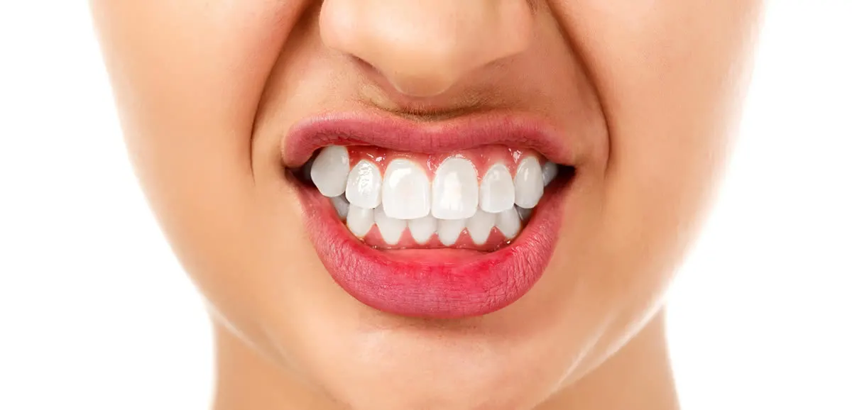
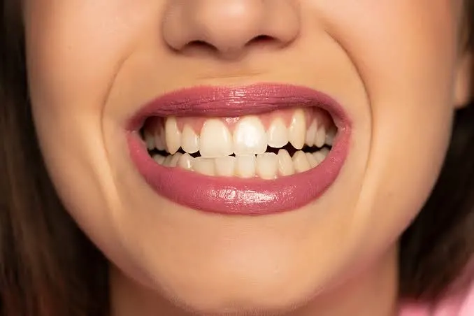
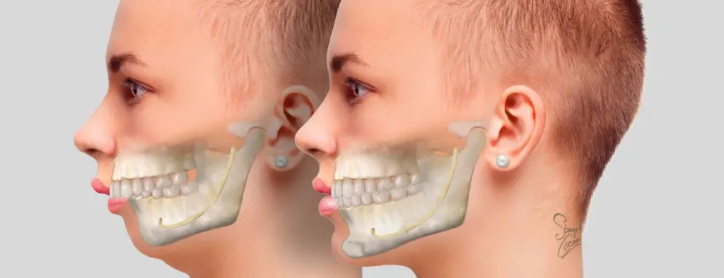
Orthognathic surgery is the surgical repositioning of not only the teeth but also the jawbones (maxilla and mandible) that support the teeth. This procedure is generally performed after skeletal growth is complete (17-18 years for women and 18-21 years for men). The primary goal of the surgery is to correct skeletal anomalies such as Class II (mandibular overbite) or Class III (mandibular overbite) to achieve an ideal Class I bite.
The process is not a standalone surgery, but a long-term treatment plan consisting of the “Orthodontics – Surgery – Orthodontics” trilogy. Surgical intervention is the most critical phase of this process, reshaping the anatomical structure.
Preoperative Preparation and Orthodontic Decompensation
When the decision for surgery is made, the patient is not taken directly to the surgery. First, a preparation process called “Pre-Surgical Orthodontics” begins. This phase usually lasts between 6 and 18 months.
What is Decompensation?
In individuals with skeletal deformities, the teeth naturally tilt at incorrect angles to mask (camouflage) the deformity. For example, in a patient with a protruding lower jaw, the lower incisors tilt back, while the upper incisors tilt forward, attempting to close the gap.
The goal of preoperative orthodontic treatment is to eliminate this natural camouflage (decompensation). The teeth are brought to their ideal angles in the jawbone. During this process, the patient’s appearance may become temporarily “disturbed” as the masking disappears and the true skeletal difference becomes apparent. This allows the surgeon to clearly see how much bone movement is required.
Digital Surgical Planning (DSP) and Simulation
Traditional methods, where planning is done on plaster models, have now been replaced by advanced technology. A detailed planning is performed immediately before surgery (while the braces are still on the teeth):
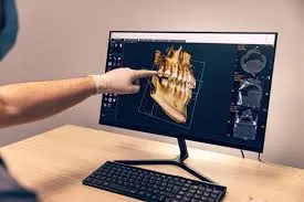
- CBCT (Cone Beam Computed Tomography): A 3D, one-to-one model of the patient’s head is created digitally.
- Virtual Surgical Planning (DSP): The surgeon virtually cuts and moves the bones on the computer. Nerve location, bone thickness, and changes in the airway are calculated in millimeters.
- Surgical Splint (Guide) Production: Acrylic guide plates (splints) are produced using 3D printers to transfer the new jaw position determined in the virtual planning to the patient during surgery. These splints are placed between the teeth during surgery, ensuring the jaws are fixed in the correct position.
Anesthesia and Introduction to Surgery
Orthognathic surgery is performed in a fully equipped operating room under general anesthesia. The anesthesia protocol requires nasotracheal intubation (insertion of a breathing tube through the nose). Oral intubation (the standard method) obstructs the surgical field because the surgeon will be working in the mouth and the teeth will be locked together.
After the patient is anesthetized, the surgical field is sterilized, and local anesthesia (with a vasoconstrictor) is administered. This is to reduce bleeding and increase postoperative comfort.
Surgical Techniques and Application Steps
In orthognathic surgery, all incisions are made inside the mouth (Intraoral Incision). This eliminates any visible scars on the face. The two most commonly used basic osteotomy (bone incision) techniques are:
A. Le Fort I Osteotomy (Upper Jaw Surgery)
This is the procedure for moving the upper jaw (maxilla).
- Incision: A horizontal incision is made under the upper lip, above the gum.
- Bone Incision: The maxilla is cut horizontally at the level of the base of the nose, using special surgical saws (piezosurgery or reciprocating saws).
- Mobilization: The upper jaw is completely separated from the skull base (down-fracture).
- Positioning: The jaw is moved to the planned new position (forward, backward, upward, or downward). If the upper jaw is to be expanded, a partial osteotomy may also be performed at this stage.
B. BSSO (Bilateral Sagittal Split Osteotomy – Lower Jaw Surgery)
This is the procedure for moving the lower jaw (mandible). It is one of the most delicate techniques because the lower jaw nerve (Inferior Alveolar Nerve) passes through this bone.
- Incision: An incision is made at the back of the lower jaw, between the cheek and gum.
- Bone Split: The jawbone is cut longitudinally in the sagittal plane using a special technique from the back (ramus). The bone is split into two pieces, like a sandwich loaf.
- Sliding: The tooth-bearing portion of the bone is shifted forward or backward, while preserving the bone with the nerve passing through it.The bone sheets slide over each other, maintaining the contact surface, which accelerates healing.
C. Genioplasty (Chin Tip Aesthetics)
When necessary, the tip of the lower jaw (mentum) is cut horizontally and moved forward, backward, or downward to balance the facial profile.
Fixation and Closure
After the bones are brought into their new position, fixation is performed to maintain this position and allow the bones to fuse (osteosynthesis).
- Rigid Fixation: The bones are firmly attached to each other using titanium plates and screws. These plates usually remain in place for life and do not need to be removed. Because they are biocompatible, the body doesn’t react.
- Intermaxillary Fixation (IMF): During surgery, the upper and lower teeth are temporarily connected to ensure the jaws are in correct occlusion. A 3D-printed splint (guide) is placed between the teeth at this stage.
- Sutures: The incisions are closed with self-dissolving (resorbable) sutures. No suture removal is required.
The First 24 Hours After Surgery and Hospitalization
The duration of surgery can vary from 2 to 5 hours depending on the extent of the procedure (single jaw or double jaw). The patient is observed in the recovery room for a period of time before being transferred to the ward.
- Ice Application (Hilotherm): Masks or ice packs that continuously apply cold are applied to the face to reduce swelling.
- Pain Management: Pain is controlled with intravenous analgesics. Patients generally report a feeling of “tightness” and “fullness” rather than pain.
- Nutrition: Only clear liquids (water, apple juice) are given for the first day.
- Sleeping Position: The head is elevated 30-45 degrees to reduce swelling.
The hospital stay is usually one or two nights.
Post-Discharge Recovery and Care
The recovery process requires patience. The body’s bone healing and soft tissue adaptation take time.
| Period | Expected Conditions | What to Do |
|---|---|---|
| Week 1 | Edema is at its highest (peaks on day 3-4). Slight bleeding may occur. | Liquid diet, regular medication use, oral hygiene (gargling), and sleeping with the head elevated. |
| Weeks 2-4 | Edema decreases by 60-70%. Rubber bands are used. Mouth opening is limited. | Transition to a soft (pure) diet. Placement and removal of elastics as directed by the dentist. |
| Week 6 | Bone union (clinical union) is largely complete. | Attempts to transition to normal (but not too hard) foods. Orthodontist follow-up. |
| Month 6 | The bone is completely healed. Edema is completely gone. | Removal of wires (by orthodontist’s decision). |
Critical Nutrition Protocol
One of the most important issues after orthognathic surgery is nutrition. Chewing forces on the jaw can disrupt the stability of plates and screws.
Diet Guidelines
First Week: A completely liquid diet. Solid soups, protein shakes, fruit juices, ayran. Using a straw is not recommended because it creates a vacuum effect and can strain the sutures; a spoon or glass should be used.
Weeks 2-4: Soft foods that do not require chewing. Mashed potatoes, yogurt, pudding, blended foods.
After Week 4: Soft chewing (pasta, omelets) begins with the physician’s approval. Tough, fibrous meats, and nuts should be avoided for at least 3 months.
Conclusion
Orthognathic surgery is a sophisticated procedure based on scientific principles, combining technology and surgical experience. The process requires the patient’s active participation and compliance. The patient’s discipline before and after surgery, as well as the technical success of the surgery, determines the longevity of the results. Correcting the skeletal structure not only provides an aesthetic improvement but also provides lifelong comfort in breathing, chewing, and speech functions.