What is Apical Resection?
The primary goal in dental practice is always to preserve the natural tooth in the mouth and maintain its function. The first treatment option for teeth infected due to decay or trauma is usually root canal treatment. However, due to the anatomical complexity of root canals, the presence of resistant bacteria, or cystic lesions at the root tip, standard root canal treatments may not always completely eliminate the infection. At this point, the surgical procedure that comes into play to save the tooth from extraction is called “Apical Resection” (Root Tip Surgery / Apisectomy). Apical resection is the surgical removal of the root tip of the tooth and the surrounding infected tissue. This procedure is a delicate and tooth-saving intervention that lies at the intersection of endodontics (the science of root canal treatment) and surgery.
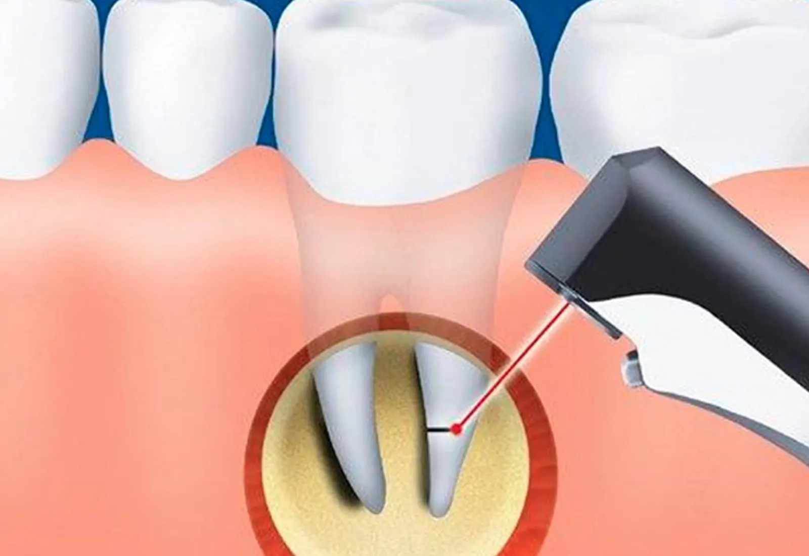
The apex of the tooth roots is a critical gateway where the vascular and nerve bundle enters the tooth, and also where infections can spread to the bone tissue. Even after root canal treatment cleans and fills the inside of the tooth, bacteria or cystic structures can sometimes persist at the root tip. This manifests as chronic pain, swelling, or the formation of a pimple-like fistula on the gum. Apical resection allows direct access to this infection focus, which is inaccessible through the root canal, by creating a window in the gum and bone. In this way, not only the infected tissue but also the root tip (usually a 3 mm portion) where the infection can reside is cut and removed. Thus, the aim is to allow the surrounding tissues to heal and the bone to regenerate without the need for tooth extraction.
Why is it done? Indications and Necessities
Apical resection is not performed after every failed root canal treatment; first, a root canal retreatment is attempted. However, in some specific cases, surgical intervention becomes the only and necessary option. The most common indication for apical resection is the presence of pathological formations called “Radicular Cyst” or “Granuloma” at the root tip. These structures are the body’s defense walls against infection, but over time they can grow and erode the jawbone. Root canal treatment alone may not always eliminate the epithelial tissue within these cysts; in such cases, surgical scraping of the cyst is necessary.
Another important cause is abnormalities in root anatomy. Some teeth may have excessively curved (dilacerated) root tips, or root canals may become blocked by calcification. In such cases, it is physically impossible to reach and clean the root tip with root canal instruments. Apical resection removes this inaccessible area by cutting the root tip. This surgical method is also used to remove foreign objects from the area in cases of complications such as filling material overflowing from the root tip during root canal treatment or the root canal instrument breaking inside the root.
Surgical Procedure: Step-by-Step Process
Apical resection is generally performed under local anesthesia and is a painless and comfortable procedure for the patient. The operation time can vary between 30 and 90 minutes depending on the position of the tooth and the size of the lesion. The procedure consists of the following steps:
In the first step, the gum tissue (gingiva) around the affected tooth is surgically removed (flap operation). Once the gum is removed, the jawbone above the root tip becomes visible. Using specialized instruments, the surgeon creates a small window in the bone and accesses the infected tissue at the root tip. The cyst or inflamed tissue (granulation tissue) is meticulously removed from the bone (curettage). Then, approximately 3 millimeters of the root tip containing the infection is cut and removed. This cutting process removes the lateral canals and microscopic bacterial nests at the root tip.
After the root tip is cut, the end of the remaining root canal is sealed with special biocompatible filling materials (MTA, Super EBA, etc.). This is called “Retrograde Filling.” This procedure is a sealing process to prevent bacteria from leaking back into the bone from the root canal. Finally, the opened bone cavity is cleaned, the removed gum tissue is placed back in its original position, and the area is closed with sutures. The healing process then begins.
Differences Between Root Canal Treatment and Apical Resection
Patients often confuse these two procedures. Root canal treatment is a cleaning done “from the inside” of the tooth, while apical resection is a procedure done “from the outside and the root tip” of the tooth. The following table summarizes the key differences between the two procedures:
g>The inner cavity of the tooth (pulp chamber).The outside of the tooth and surrounding bone tissue.
| Feature | Root Canal Treatment (Endodontics) | Apical Resection (Surgery) |
|---|---|---|
| Access Route | From the chewing surface of the tooth (Crown). | Through the gum and bone (From the root tip). |
| Purpose | To disinfect the root canal. | To scrape away the cyst/infection at the root tip. |
| Area of Application | ||
| Healing | No surgical wound, heals immediately. | Healing of a sutured wound (7-10 days). |
| Timing | It is the first treatment of infection. | It is performed in cases where root canal treatment has failed. |
Postoperative Recovery and Important Considerations
The healing process after apical resection is generally smooth and fast. However, since it is a surgical procedure, there are some things to consider in the first few days. Slight swelling (edema) and bruising may be seen on the face immediately after the procedure; this is a normal and expected occurrence. Applying ice compresses (cold application) externally for the first 24 hours is very important to minimize swelling. Regular use of antibiotics, painkillers, and antiseptic mouthwashes prescribed by the doctor eliminates the risk of infection.
Nutrition: Avoid hot, hard, and spicy foods in the first few days; warm and soft foods should be preferred.
Oral Hygiene: Do not brush the area where the stitches are located, but continue cleaning the other teeth. The wound area should be kept clean with the mouthwashes recommended by the doctor.
- Smoking and Alcohol: Smoking and alcohol should not be consumed for at least 3-5 days after the operation as they delay healing and impair blood flow.
Conclusion
In conclusion; apical resection is one of the most important applications of the tooth preservation philosophy of modern dentistry. In cases of persistent infections, cysts, or anatomical difficulties, instead of discarding the tooth, it’s possible to give it a second chance with a surgical intervention. When performed with the correct indications and by expert hands, this procedure has a very high success rate, allowing the patient to continue using their own natural tooth. Remember, no prosthesis or implant can completely replace a person’s own natural tooth biologically and functionally.