What are the Stages of Implant Treatment in Kahramanmaraş?
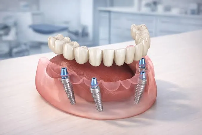
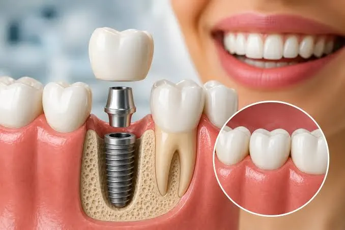
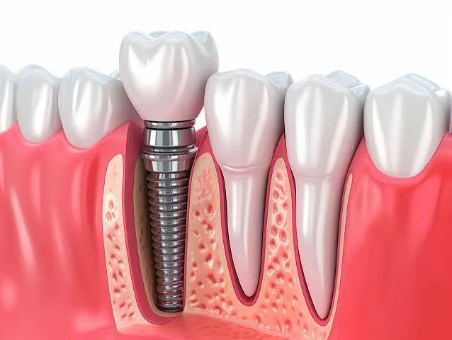
In human anatomy, the mouth and jaw structure is a highly complex system that, beyond being the starting point of the nutritional function, directly affects the individual’s psychosocial life in terms of speech (phonation) and aesthetic integrity. Tooth loss experienced due to cavities, traumas, or progressive periodontal (gum) diseases causes chain reactions in this flawless biomechanical system. The root cavity of a lost tooth eventually leads to the melting of the jawbone in that area (bone resorption), the tipping of adjacent teeth towards this gap, and the complete disruption of the mutual jaw closure balance. While traditional dentistry applications try to eliminate these gaps with bridge prostheses where adjacent teeth are cut and reduced, the gold standard of today’s modern medical world is “dental implant” treatments. Implants are high-tech artificial roots produced from tissue-friendly (biocompatible) titanium material that mimic the root of the lost tooth at a biological level. For patients seeking an answer to the question **what are the stages of implant treatment in Kahramanmaraş**, it is of great importance to know that this process is not just a screw placement procedure; it entails serious radiological analysis, digital engineering, and a cellular healing phase behind it.
Biological Infrastructure: The Harmony of the Implant and Jawbone
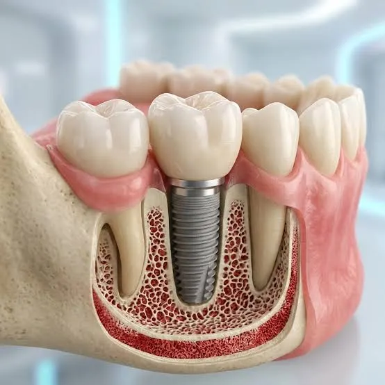
Before moving on to the clinical stages of the treatment, it is necessary to understand the biological mechanism that forms the basic philosophy and success criterion of implantology. This phenomenon, called “osseointegration” in medical literature, is the process of the titanium surface being accepted by living jawbone cells (osteoblasts) and being tightly embraced at a microscopic level. Titanium is one of the most biocompatible elements in nature, which the human immune system does not perceive and reject as a foreign body. The surface of the implant is roughened with special acids or sandblasting techniques during the production phase so that bone cells can attach faster and stronger (hydrophilic surface technologies).
Dentocare Klinik, serving in the Kahramanmaraş location, starts the process by selecting implant systems with international quality standards according to the patients’ bone density, anatomical structure, and the type of prosthesis to be applied. A successful osseointegration process forms the basis of the resistance the implant will show against chewing forces in the coming years. Therefore, the angle of placement of the implant into the bone, the torque (compression) force applied to the tissue, and the thickness of the surrounding gum (biotype) are the works of not only a mechanical but also a biological harmony.
Stage 1: Digital Diagnostics and 3D Planning (Preparation Phase)
While operations in traditional dentistry are often planned over two-dimensional panoramic x-rays, modern implant surgery has completely turned into a three-dimensional (3D) engineering work thanks to the innovations brought by the digital age. In this first and most critical stage of the treatment process, the patient’s general systemic history (conditions such as diabetes, hypertension, osteoporosis) is listened to in detail. This is because cellular healing capacity is directly linked to general body health.
Then, a three-dimensional map of the patient’s jawbone is created with the 3D Volumetric Dental Tomography (CBCT) device available in the clinic. Thanks to this scan, the millimetric height, thickness, quality of the spongy (cancellous) or hard (cortical) structure of the jawbone, the position of the nerve canals passing through the lower jaw (nervus alveolaris inferior), and the borders of the sinus cavities in the upper jaw are perfectly determined. Dentocare Klinik experts transfer these digital data to special software and perform the surgical operation virtually on the computer screen. Which diameter and length of implant will be placed, at what angle, and exactly where, is determined days in advance. In deemed necessary cases, this virtual planning is converted into “surgical guide plates” with 3D printers, guiding the physician during the operation and reducing the margin of error to zero.
Stage 2: Minimally Invasive Surgical Intervention (Placement of the Implant)
The surgical stage, where digital planning is reflected in the clinic, is a procedure that patients are usually most anxious about, but thanks to local anesthesia, it is completely painless and takes a very short time. The area to be treated is numbed under high sterilization protocols. In “digitally guided” operations where bone volume and gum thickness are suitable, the procedure can be performed without cutting the gum with a scalpel and separating it from the bone (flapless surgery / seamless implant technique), by opening a socket only as small as the diameter of the implant. This minimally invasive (least damaging to the tissue) approach minimizes the edema (swelling) and discomfort that may occur after the operation, significantly speeding up the patient’s return to social life.
The implant socket is prepared precisely using gradually expanding sterile titanium burs on the jawbone. In order to prevent bone cells from being damaged by high heat (thermal necrosis) during this process, the area is continuously cooled with sterile saline. After the socket is prepared, the titanium implant is slowly placed into the bone with a pre-targeted torque value. If the implant holds onto the bone tightly enough (high primary stability), the healing cap that will shape the gum can be attached immediately. In cases where the bone is weak or bone powder (grafting) is added during the procedure, the implant is left under the gum, sutured sterilely, and completely retreats into a biological seclusion.
Clinical Stages and Process Timeline
The adventure of a dental implant integrating with the jawbone and turning into a final tooth requires respecting the rhythm of tissue healing. Although each patient’s metabolic rate is different, the biological calendar of the basic treatment phases applied at Dentocare Klinik standards is summarized in the table below:
| Clinical Treatment Phase | Estimated Duration and Waiting | Basic Biological and Medical Goals |
|---|---|---|
| Radiological Diagnosis and Planning | 1-2 Days | Resetting anatomical risks to zero with 3D tomography, measurement of bone quality, and surgical guide preparation. |
| Surgical Implantation (Operation) | 30 – 60 Minutes (Depending on the Number of Implants) | Placing the titanium root into the jawbone at an ideal angle by keeping tissue trauma to a minimum (Primary Stability). |
| Osseointegration (Healing) Phase | Lower Jaw: 2-3 Months | Upper Jaw: 3-4 Months | Fusion of bone cells (osteoblasts) to the implant at a microscopic level and ensuring mechanical integrity. |
| Prosthetic Loading and Aesthetic Finish | 1-2 Weeks (From Impression to Delivery) | Gingival (gum) shaping, digital intraoral measurement, abutment adaptation, and zirconium/porcelain crown integration. |
Stage 3: Osseointegration and Biological Waiting
The process that starts after getting up from the operation chair is the period when medical technology shakes hands with the body’s miracle of self-repair. In this cellular fusion phase, which we call osseointegration, since the lower jaw bone is structurally denser (cortical), the integration process generally takes 2 to 3 months; whereas, since the upper jaw bone has a more porous (cancellous) structure, this waiting period can take up to 3 to 4 months. If “Sinus Lifting” (lifting the sinus membrane) or horizontal/vertical bone powder addition (augmentation) is performed during the surgery, this period can be extended by a few more months for the tissue to mature.
To ensure that this waiting phase does not turn into an aesthetic and functional loss for patients, custom-made temporary prosthesis alternatives (removable or implant-supported fixed temporaries) are applied by Dentocare Klinik. Thus, while the bone cells do their job, patients can continue their daily social lives and speech comfort without interruption. The biggest factor that disrupts tissue blood supply and hinders healing during this period is the use of tobacco. For a successful fusion process, limiting or completely quitting smoking is of great importance for the health of the process.
Stage 4: Digital Impression, Abutment, and Prosthetic Loading
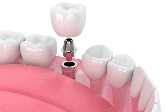
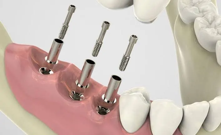
When that strong bond between the bone and the titanium artificial root is established, the most enjoyable and aesthetic stage of the process, where the result is directly felt by the patients, begins. If the implant was hidden under the gum during the operation, it is uncovered with a laser or a small cautery touch, and “healing caps” are attached to make the gum suitable for the emergence profile of a natural tooth. After the gum achieves the desired smooth and circular form, the impression stage for the construction of the crown (upper tooth) is reached.
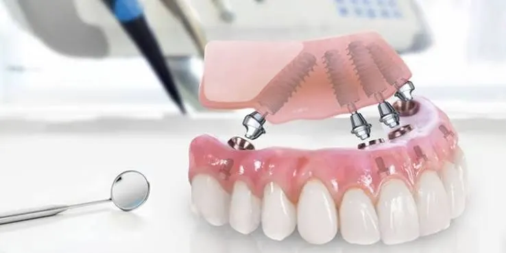
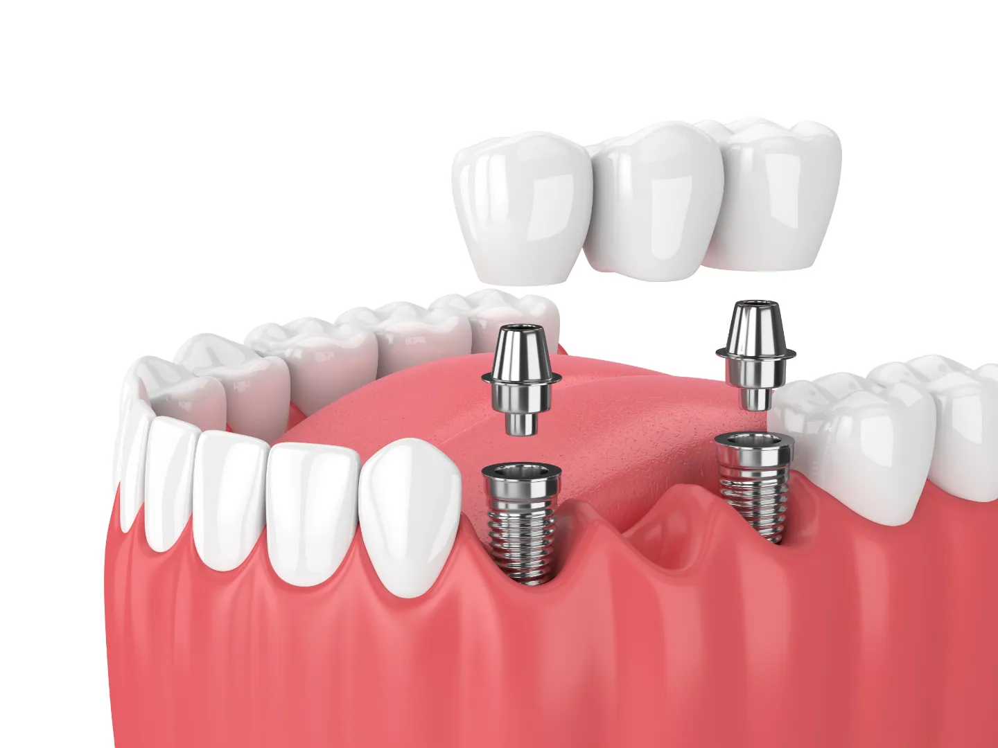
In today’s digital dentistry, traditional impression pastes that cause nausea are largely abandoned. At Dentocare Klinik, millimetric 3D photos of the teeth are taken in seconds with intraoral scanner cameras and transferred to the digital environment. On these digital models, the intermediate piece (abutment) to be connected to the implant and the final upper prosthesis (zirconium or all-ceramic crowns) are designed with zero margin of error using CAD/CAM technologies. Thanks to the light transmittance of zirconium and its tremendous tissue compatibility with the gum, your new implanted tooth gains an optical depth and chewing durability indistinguishable from your neighboring natural teeth.
Two Golden Rules for Long-Lasting Implant Health
Although this new structure mechanically and biologically integrated into your mouth does not carry a risk of cavities; the gum and bone tissue surrounding the implant must be protected against microbial infections (peri-implantitis). To keep this unique investment healthy for years, the most basic approaches you need to integrate into your daily routine are:
- Specific Interdental Hygiene: The standard tooth brushing procedure may be insufficient to clean the plaques at the implant and gum junction. To prevent food accumulation and gingivitis in this area, the regular use of water flosser devices, under-bridge floss (superfloss), or interdental brushes is absolutely essential.
- Regular Radiological Follow-up: The intraoral environment is dynamic. In order to intervene before any infection reaches the bone, having panoramic x-ray supported physician check-ups at Dentocare Klinik in 6-month periods is the greatest insurance that extends the life of your implant.
Conclusion: The Strongest Investment Made in Your Health and Future
To summarize, the question of what are the stages of implant treatment in Kahramanmaraş is a summary of a multidisciplinary medical story where cellular healing, high engineering, and clinical experience come together. Living with tooth deficiencies disrupts your chewing habits and thus your stomach/digestive health; whereas, modern dental implants provide the closest mechanical support to the original of nature, revealing satisfying results.