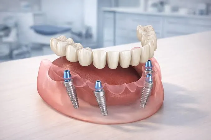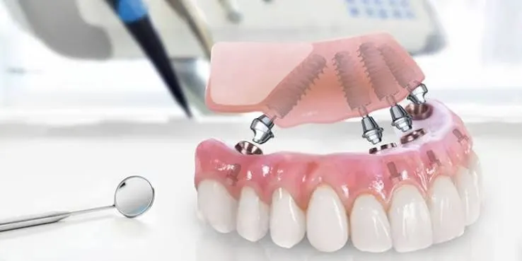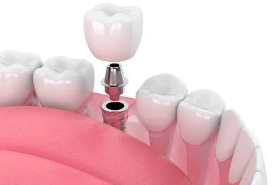
Kahramanmaraş Implant Diagnosis
A Zero-Risk Goal in Maxillofacial Surgery in the Light of 3D Tomography, Systemic Analysis, and Scientific Data at Dentocare Clinic
The Foundation of Successful Surgery: Accurate and Detailed Diagnosis
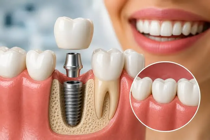
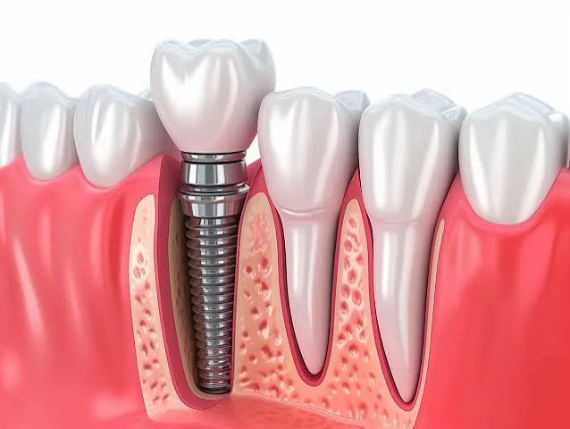
The most fundamental rule widely accepted in medical science is this: “The right treatment can only be built on a flawless diagnosis.” When it comes to replacing missing teeth and placing a titanium material into the jawbone, the diagnostic stage goes beyond a simple dental check-up and becomes a multidisciplinary process requiring advanced radiological imaging and systemic evaluation. In Kahramanmaraş implant diagnosis applications, Dentocare Clinic begins treatment by analyzing the anatomical structures of its patients not through rough estimations, but through millimetric digital data. This is because the density, quality, and anatomical relationships of the jawbone are as unique to each individual as a fingerprint.
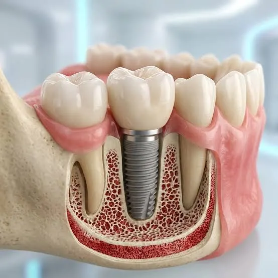
In the areas where teeth have been lost, bone resorption (atrophy) begins over time as a physiological process. A bone structure that may appear sufficient from the outside or on standard two-dimensional X-rays may, in fact, not have enough internal volume to support an implant. Dentocare Clinic specialists aim not only for the implant to attach to the bone, but also to withstand chewing forces for decades. In line with this goal, the diagnostic protocols applied are designed to resolve all anatomical surprises that may be encountered during surgery in advance and to provide the patient with a safe medical roadmap.
Multidimensional Diagnostic Protocols: Step-by-Step Analysis
Every patient who applies to our clinic requesting implant treatment goes through the following diagnostic stages, which maximize safety and fully comply with medical ethics:
- Systemic Health Anamnesis
The most important factor affecting cellular healing (osseointegration) in the jawbone is overall body health. Our dentists thoroughly evaluate systemic conditions such as uncontrolled diabetes, osteoporosis, hypertension, or the regular use of blood thinners/bone medications. When necessary, consultation is made with your medical doctors (cardiologist, endocrinologist), and approval for surgery is obtained. - Clinical Intraoral Examination
The missing tooth area is physically examined. The width of the extraction site, the biotype (thickness) of the gum tissue, the root angles of adjacent teeth, and the occlusal relationship between the jaws are evaluated. - 3D Volumetric Dental Tomography (CBCT)
This is the heart of the diagnosis. Unlike 2D images, your jawbone is digitally examined in a full 360-degree rotation. Bone volume, width, and height are measured with millimetric precision. The nerve canal in the lower jaw that provides sensation to the lip (Nervus Alveolaris Inferior) and the sinus cavities in the upper jaw are mapped with certainty. - Bone Quality Assessment (D1-D4 Scale)
It is not enough for the bone merely to be present; its quality is also important. Through tomography data, it is determined how hard or spongy your bone is (from D1 to D4). This data allows us to select the surface characteristics and thread design of the implant to be used.
The Role of Imaging Technologies in Diagnosis
Our patients often wonder about the difference between a panoramic X-ray and tomography. You can review in the table below how accurate diagnosis in maxillofacial surgery depends on radiological depth:
| Diagnostic Tool | Panoramic X-Ray (2D) | Dental Tomography – CBCT (3D) |
|---|---|---|
| Image Dimension | Reflects the bone only in terms of height and width (two-dimensional). | Provides three-dimensional (volumetric) data that also shows the depth (thickness) of the bone. |
| Bone Density | Does not provide definitive information about the hardness (quality) of the bone. | Measures the internal density of the bone with millimetric precision using Hounsfield units. |
| Anatomical Precision | Structures such as nerves and sinuses may be misleading due to overlapping shadows. | Thanks to cross-sectional scanning, nerve canals and sinuses are mapped with zero margin of error. |
| Field of Application | Sufficient for general dentistry diagnoses (caries, impacted wisdom teeth, etc.). | The gold standard in advanced maxillofacial surgery planning such as implants. |
Post-Diagnosis Case Management and Grafting (Bone Powder)
If insufficiency is detected in the jawbone as a result of the detailed three-dimensional diagnosis, this does not mean that implant treatment will be abandoned. On the contrary, identifying this deficiency before surgery makes it possible to plan “infrastructure reinforcement” procedures on time. In cases where bone height or thickness is insufficient, Dentocare Clinic specialists include “bone grafting” (bone powder addition) or “sinus lifting” operations in the diagnostic plan, thereby building a solid biological foundation on which the implant can be safely placed.