Tooth Extraction Diagnosis in Kahramanmaraş
In dentistry, the most irreversible and non-recoverable procedure is the permanent removal of a tooth from the jawbone. For this reason, deciding to extract a tooth can never be a superficial judgment based solely on the pain the patient feels or on a cavity that is visible to the eye. The heart of proper treatment planning is a flawless diagnostic process grounded in scientific data, radiological evidence, and specific clinical tests. Dentocare Clinic, a pioneer of advanced dental practices in the Kahramanmaraş region, transforms tooth extraction diagnosis from a simple assumption into an analytical process managed according to the principles of evidence-based medicine.
Many patients who come to the clinic with severe pain lasting for days assume that the source of that pain must certainly be a decayed tooth that needs to be extracted. However, the anatomy of the jaw and face contains an extremely complex nerve network. Sometimes the source of pain may be a neighboring tooth, sometimes an inflammation in the sinus cavity (sinusitis), and sometimes the temporomandibular joint (TMJ) or a spasm in the chewing muscles. If the correct diagnosis is not established, a completely healthy tooth may be extracted by mistake while the patient’s actual pain continues. To reduce these risks to zero, Dentocare Clinic’s specialist dentists activate differential diagnosis protocols and aim to identify the true biological and pathological source of the pain with absolute accuracy.
From Symptom to Diagnosis: Scientifically Reading the Character of Pain
The first source of information our dentists rely on in tooth extraction diagnosis is the neurological character of the pain experienced by the patient. The pulp, the living tissue inside the tooth containing nerves and blood vessels, reacts differently depending on the depth of the infection. A short-lasting ache that occurs only when drinking cold water usually indicates a “reversible” damage that can often be saved with a filling or root canal treatment. On the other hand, throbbing pain that wakes the patient at night, increases with heat, and does not respond to painkillers suggests that the dental nerve has completely died (necrosis) and that the infection has spread into the jawbone.
After listening to these symptoms, Dentocare Clinic dentists subject the tooth to physical and chemical tests to confirm the diagnosis. The level of infection in the tooth, the destruction in the jawbone, and the effect on surrounding tissues are converted into measurable clinical data through these specific tests.
Clinical Tests and Methods That Confirm the Extraction Decision
At our clinic, a visible cavity alone is never enough to place a tooth on the extraction list. The main clinical evaluation steps that prove the tooth is a hopeless case and clarify the diagnosis are as follows:
- Vitality Tests: Special cooling sprays or digital electric pulp test devices (EPT) are applied to the tooth to determine whether the dental nerve is still alive. If the tooth does not respond to any stimulus, the tissue has lost its vitality and is considered necrotic.
- Percussion and Palpation: Our dentist gently taps the tooth vertically and horizontally with the handle of a dental mirror (percussion) to test whether there is an infection in the periodontal ligament around the root tip. Pressure applied by hand to the gum in line with the root (palpation) helps diagnose the outward manifestation of a cyst or abscess within the bone.
- Periodontal Probing: The depth of the pocket between the tooth and the gum is measured with special millimetric instruments. If the probe descends very deep into the jawbone along the root of the tooth (for example, 8–10 mm), this medically proves that the bone holding the tooth has completely resorbed and that the tooth has no chance other than extraction.
Radiological Evidence: 2D vs. 3D in Extraction Diagnosis
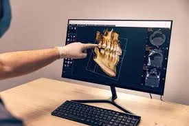
If clinical tests represent half of the diagnosis, radiological images represent the other half. Bone loss around the root of a tooth appears on X-rays as radiolucency—dark areas indicating where the bone has dissolved. The accuracy of the diagnosis is directly proportional to the quality of the device being used. In the table below, you can see the place of imaging methods used in diagnosis within Dentocare Clinic’s infrastructure:
| Traditional (2D) Diagnostic Approach | Dentocare (3D CBCT) Diagnostic Approach |
|---|---|
| On panoramic X-rays, bone thickness cannot be clearly understood, and because cysts or roots may overlap one another (superposition), a misleading diagnosis may be made. | With dental tomography, the tooth root is examined in millimetric slices, there is no shadow overlap, and the true volume of the root-end lesion can be calculated with complete clarity. |
| In impacted wisdom teeth, the exact distance to the inferior alveolar nerve in the lower jaw can only be estimated, since there is no depth perception. | The relationship between the nerve canal and the tooth root—whether it lies to the right or left and how close it is—is mapped in a 3D environment, enabling a zero-risk preoperative extraction diagnosis. |
| Vertical root fractures are often missed on 2D films, and unnecessary root canal treatments may be attempted on the tooth. | Even the finest hairline root fractures are detected immediately on tomography, allowing a direct extraction and implant decision without wasting the patient’s time or money. |
From Diagnosis to Treatment: Dentocare Clinic’s Transparent Roadmap
Once all of these clinical and radiological data have been gathered, the “diagnosis” regarding the condition of your tooth ceases to be an assumption and becomes a scientific reality. Dentocare Clinic specialists display this diagnosis on patient monitors and explain step by step, in visual form, the bone loss in your jaw, the loss of tooth substance, or the cystic structure. When you understand the logical and medical basis of why a tooth needs to be extracted, the uncertainty and fear you feel toward the procedure naturally disappear.
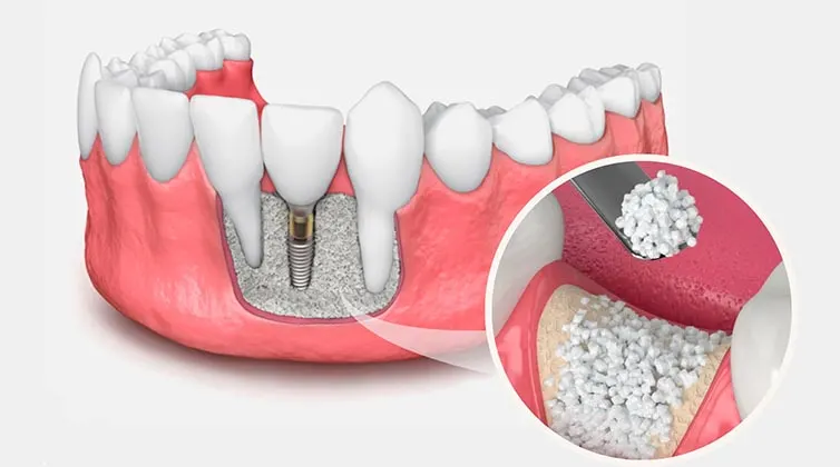
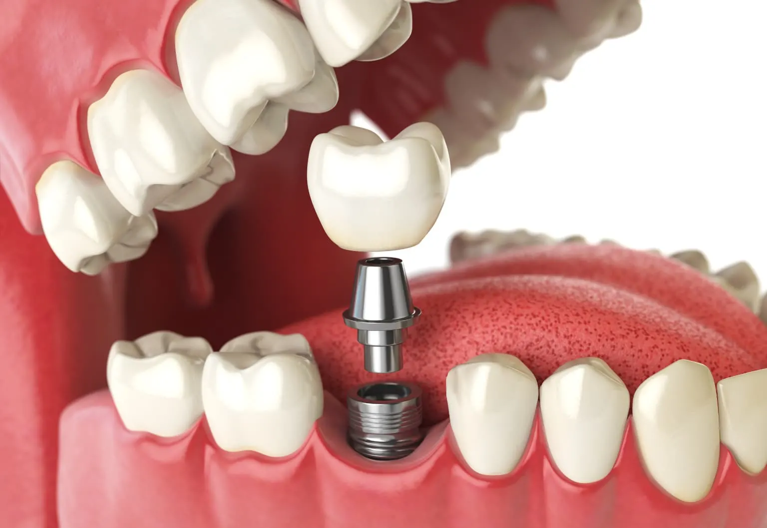
Once the diagnosis is clarified, the process no longer focuses only on pulling the tooth. It evolves into a broad treatment plan centered on how that area will be rehabilitated after extraction, including socket preservation, same-day implant placement, or bridge prosthetics. Because the correct diagnosis aims not only to stop today’s infection, but also to secure your future chewing health.
Contact Us for Scientific Diagnosis and Proper Guidance
Oral and jaw health is far too valuable to be managed through assumptions or trial-and-error methods. The fate of a painful or loose tooth should only be determined by a flawless diagnostic process supported by advanced technology and filtered through the analytical judgment of an expert dentist. An incorrect diagnosis may cost you a healthy tooth, or allow an unnoticed infection to silently dissolve your jawbone.
If you have serious doubts about your dental health in Kahramanmaraş and the surrounding area, and if you are looking for an evidence-based, scientific, and honest answer to the question, “Does this tooth really need to be extracted?”, then you are in the right place. Dentocare Clinic is ready to map your health with the greatest accuracy through its modern diagnostic infrastructure, 3D imaging systems, and experienced team of specialist dentists. To begin your treatment with the confidence of a true diagnosis and to schedule your detailed analysis appointment, contact our clinic today.