Kahramanmaraş Implant Planning
Millimetric Preventive Engineering with Digital 3D Tomography and Surgical Guide Technologies at Dentocare Clinic
The Secret of Success in Implantology: Perfect Pre-Planning
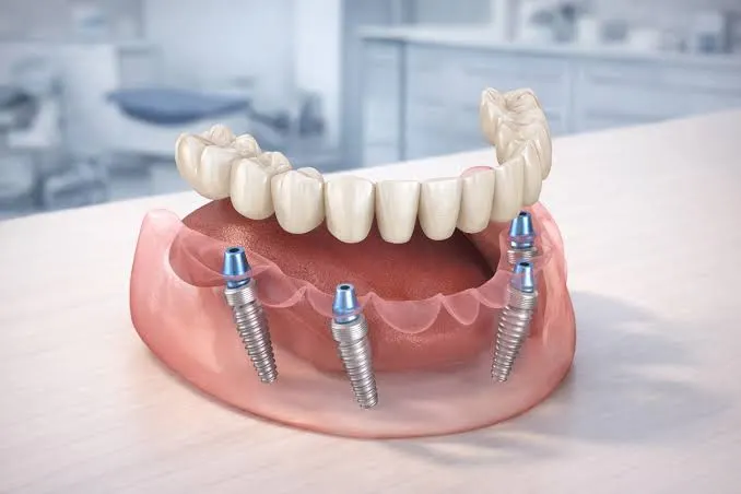
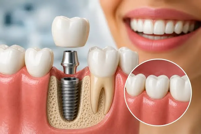
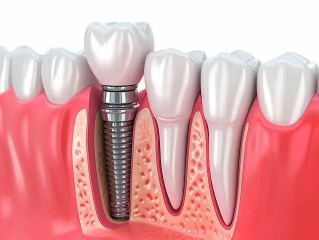
In dentistry and maxillofacial surgery, success depends not on performance during the operation, but on detailed analyses and digital engineering conducted beforehand. Placing a titanium root into the jawbone should not be like navigating in the dark, but like progressing on a clear and safe map where every millimeter has been pre-calculated. The process of creating this map is called Kahramanmaraş implant planning. At Dentocare Clinic, we do not consider implant treatment as merely a mechanical placement procedure; we approach it as a biological reconstruction project in which human anatomy, biomechanics, and digital technology work in perfect harmony.
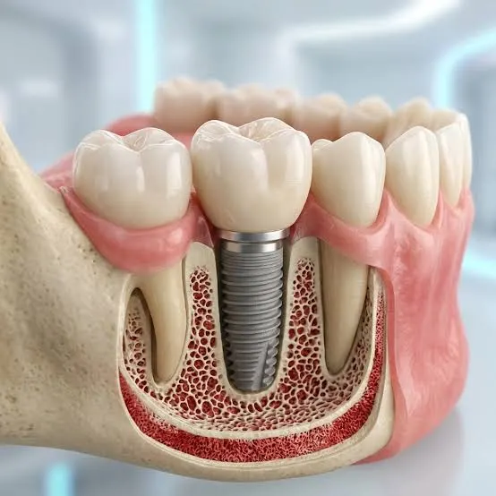
Just as architectural drawings, soil surveys, and structural calculations are carried out before laying the foundation of a building, a “ground survey” of your jawbone must also be performed before placing implants, which form the foundation of your new teeth. Your bone density, the pathways of your jaw nerves, the position of the sinus cavities in the upper jaw, and the root angles of adjacent teeth are critical parameters that determine the treatment route. The digital planning protocols applied in our clinic aim to eliminate all possible anatomical surprises during surgery (such as nerve damage or sinus perforation) and to provide the patient with a completely comfortable surgical experience.
Dentocare Clinic Digital Planning Stages
Two-dimensional panoramic X-rays, frequently used in traditional dentistry, only provide information about the height and width of the jawbone and may be misleading regarding its depth and actual volume. At Dentocare Clinic, the planning process consists of the following digital stages that eliminate the margin of error:
- 1. 3D Volumetric Dental Tomography (CBCT)
The patient’s jaw structure is scanned in three dimensions within seconds. This scan allows the jawbone to be examined in cross-sections. Areas of bone loss and the distance of nerve pathways from the implant site are measured with millimetric precision. - 2. Virtual Surgical Simulation
Tomography data are transferred to specialized software. Within this software, a virtual model of the jawbone is created. By selecting the diameter and length of the titanium implant, the dentist performs a virtual placement at the most ideal angle within the bone. The distribution of chewing forces is also calculated at this stage. - 3. Surgical Guide Production
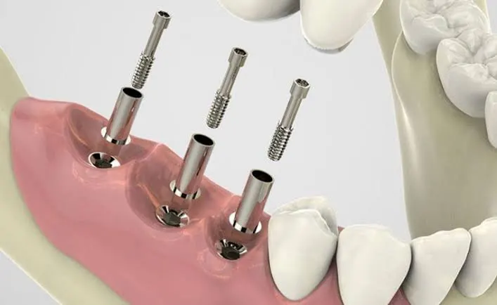
The flawless planning carried out in the virtual environment is transformed into a transparent template using 3D printers. This “surgical guide,” placed in the patient’s mouth on the day of the operation, directs the dentist’s drill only at the pre-planned angle and depth. Thus, any margin of error that could occur by human hand is anatomically eliminated. - 4. Prosthetic (Superstructure) Planning
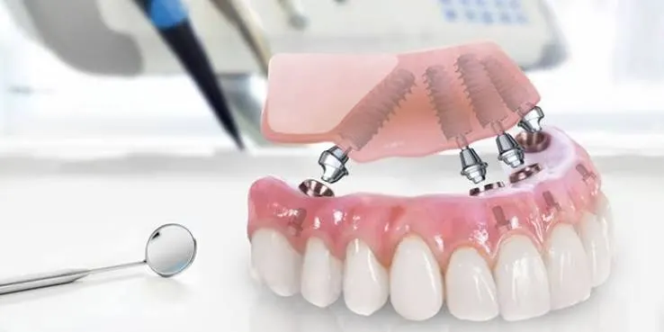
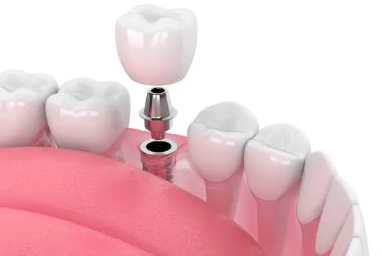
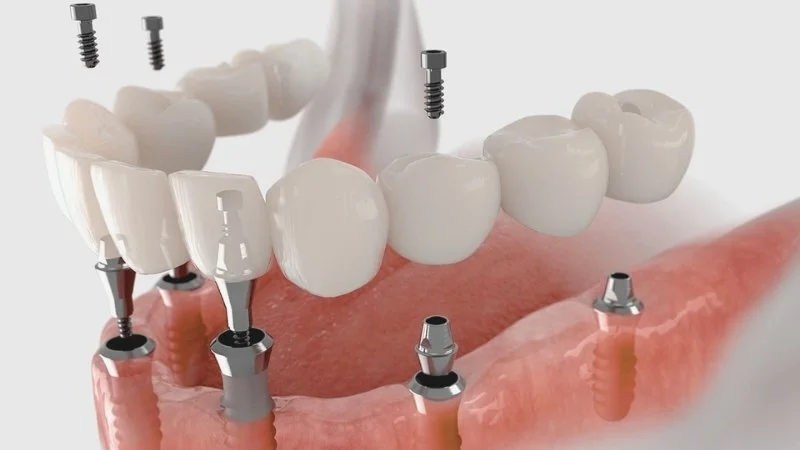
An implant is not just a screw placed into the bone; it is the foundation that determines the position of the aesthetic tooth to be placed on top. During the planning stage, how the future porcelain or zirconia tooth will align with the patient’s facial contours, smile line, and lip support is digitally designed in advance (with smile design integration).
Advantages of Digital Planning Compared to Conventional Methods
Our patients naturally wonder how this advanced technological infrastructure translates into their treatment comfort. You can clearly see the difference between our digital approach and traditional methods in implant planning in the comparison table below:
| Clinical Criteria | Dentocare Digital Planning Approach | Traditional (Routine) Clinical Approach |
|---|---|---|
| Depth of Diagnosis | Bone volume, sinus, and nerve positions are examined 360 degrees with 3D tomography. | Only the width and height of the bone are estimated using 2D panoramic X-rays. |
| Application Safety | With surgical guides, the dentist’s drill cannot exceed the predetermined limits. (Maximum Safety) | Relies on the dentist’s real-time observation, anatomical estimation, and manual experience (free-hand). |
| Surgical Trauma (Incision) | Since the exact position is known, procedures can often be performed without cutting the gum tissue (flapless/sutureless). | The gum tissue must be widely incised and opened to visualize the bone, and then sutured afterward. |
| Healing Comfort | Due to minimal tissue trauma, post-operative swelling, edema, or discomfort is almost negligible. | Due to incisions and sutures, there is a higher likelihood of post-operative swelling and sensitivity. |
The Power of Planning in Preventing Complications
The most feared scenarios in implant surgery are pressure on the nerve passing through the lower jaw (Nervus Alveolaris Inferior), causing numbness in the lip, or perforation of the sinus cavity in the upper jaw, leading to infection. Thanks to the detailed radiological and digital implant planning performed at Dentocare Clinic, such anatomical risks are completely eliminated. Before intervening in your bone structure, our dentists map out all potential risks and, if necessary, include preparatory procedures such as “bone grafting” or “sinus lifting” in your treatment plan.