Kahramanmaraş Orthognathic Surgery Diagnosis: Accurate Identification of Skeletal Problems
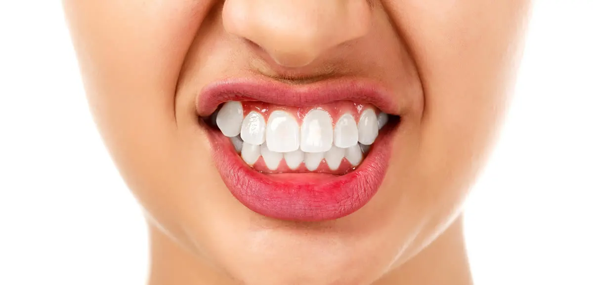
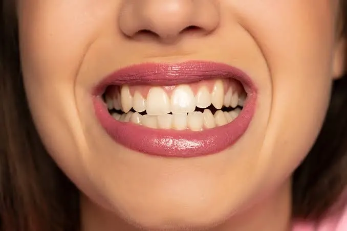
Disorders in the dental and jaw structure are often perceived from the outside as merely aesthetic issues. Patients usually present to the clinic with complaints such as “my teeth are crooked” or “my jaw looks too forward.” However, these visible complaints are actually just the external manifestation of a much deeper skeletal anomaly. This is where Kahramanmaraş orthognathic surgery diagnosis comes into play. The diagnostic phase is the art of clearly determining whether the problem is a superficial dental alignment issue or a jaw deformity directly caused by the position of the bones. As Dentocare Clinic, we carry out this diagnostic process—which determines the entire course of treatment—using evidence-based medical data and advanced technological radiological analyses.
Why Is Diagnosis the Most Critical Stage of Treatment?
There is a general rule in medicine: an incorrect diagnosis does not lead to the correct treatment. In cases requiring orthognathic surgery (jaw surgery), this rule applies even more strictly. If a patient’s lower jaw is skeletally retruded and this condition is attempted to be “camouflaged” only with braces, the result is often disappointing. Since the teeth are excessively forced to move within the bone, root resorption may begin, gum recession may occur, and shortly after treatment, the teeth may tend to relapse to their former misaligned positions.
Dentocare Clinic physicians focus on the root cause when making a diagnosis. Determining whether a problem is “dental” or “skeletal” is clarified through the joint decision of our orthodontic and maxillofacial surgery specialists. A patient diagnosed with orthognathic surgery understands that the problem is too complex to be resolved merely by repositioning the teeth, and that the upper and lower jawbones are incorrectly positioned relative to each other and to the cranial base. This clear diagnosis saves the patient from unnecessary time and financial loss, directing them straight to a definitive solution.
Cephalometric Analysis: The Mathematical Foundation of Diagnosis
A diagnosis in jaw surgery cannot be made by visual estimation or by evaluating physical appearance alone. During examination, millimetric angles are calculated on profile X-rays (cephalometric radiographs). These mathematical measurements form the scientific basis of the diagnosis. The main parameters carefully evaluated by our physicians during diagnosis include:
- SNA Angle: Determines how far forward or backward the upper jaw (maxilla) is positioned relative to the cranial base.
- SNB Angle: Indicates the position of the lower jaw (mandible) relative to the cranial base. Deviations in this angle provide clear evidence of mandibular retrusion or protrusion.
- ANB Angle: Reveals the relationship between the upper and lower jaws. The degree of skeletal discrepancy is classified based on this angle.
- Soft Tissue Profile: The harmony between the lips, cheeks, and nose (which cover the bones) and the underlying skeletal structure is analyzed. The distance of the chin to the lips (E-line) is calculated to refine the aesthetic diagnosis.
Skeletal Malocclusion Classification in Orthognathic Surgery
At Dentocare Clinic, our patients’ diagnoses are classified according to skeletal categories accepted in international medical literature. Knowing which skeletal class you belong to directly determines the scope of the operation and the treatment protocol. You can review the main anomaly classes identified during the diagnostic phase in the table below:
| Diagnosis Class | Anatomical Condition and Symptoms | Clinical Manifestation |
|---|---|---|
| Class I (Skeletal Norm) | The position of the jawbones relative to each other and to the skull is normal. The problem exists only in the teeth. | Usually does not require surgery; only orthodontic treatment (braces/aligners) is indicated. |
| Class II (Retrognathism) | The lower jaw is positioned too far back or the upper jaw is excessively forward. The ANB angle is larger than normal. | A “bird-face” profile appears. The patient cannot properly bite with the front teeth. Mandibular advancement surgery is indicated. |
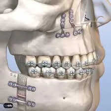
| Class III (Prognathism) | The lower jaw has grown forward beyond the upper jaw skeletally. The ANB angle is negative. | A prominent and strong chin, reverse bite (lower teeth overlapping upper teeth) are observed. Mandibular setback and/or maxillary advancement is indicated. |
| Asymmetry / Open Bite | Imbalance between the right and left sides of the jaws or a condition where the front teeth remain open while the back teeth are in contact. | The facial midline is shifted. Chewing function is severely impaired. Double jaw surgery is commonly indicated. |
Airway Narrowing and Sleep Apnea Diagnosis
There is another dimension of orthognathic diagnosis that is often overlooked but directly affects the patient’s lifespan and quality of life: airway analysis. Especially in patients diagnosed with skeletal Class II (mandibular retrusion), the tongue base is positioned backward toward the airway due to the retruded lower jaw. This anatomical narrowing causes snoring at night, pauses in breathing during sleep (Obstructive Sleep Apnea), and waking up constantly fatigued in the morning.
In the 3D tomography analyses performed at Dentocare Clinic, the patient’s airway volume is calculated volumetrically. If severe airway narrowing is diagnosed, the goal of orthognathic surgery goes beyond creating an aesthetic profile; it becomes a permanent airway expansion procedure that ensures comfortable breathing. Surgically advancing the lower jaw physically enlarges the airway and provides a radical treatment for sleep apnea syndrome.
Detection of Functional Damage: Temporomandibular Joint (TMJ) Disorders
Skeletal disorders prevent the upper and lower teeth from closing in a perfect lock-and-key relationship. Incorrect tooth contact causes the entire chewing force to be unevenly and excessively loaded onto the Temporomandibular Joint (TMJ), which connects the lower jaw to the skull. During diagnosis, not only the bone dimensions but also the health of the joint disc are evaluated.
If the patient experiences limited mouth opening, clicking sounds in the joint while eating, or chronic head and neck pain, this is recorded as a secondary diagnosis. Repositioning the jaws to their anatomically correct position through orthognathic surgery removes this pathological load on the joint and provides functional rehabilitation.
Dentocare Clinic: A Safe Future with Accurate Diagnosis
If you suspect a skeletal problem affecting your jaw and facial region in the Kahramanmaraş area, the first and most important step you should take is obtaining an accurate medical diagnosis. Jaw asymmetries and excessively forward or backward jaw structures do not resolve on their own over time; on the contrary, they may lead to irreversible wear in joint tissues as age progresses. The solution is not to postpone the problem with temporary methods, but to clearly identify its root at the skeletal level.
Our expert maxillofacial surgeons and orthodontists at Dentocare Clinic are ready to provide you with the most accurate and transparent diagnosis using detailed cephalometric analyses and the latest medical technologies. To achieve not only an aesthetic smile but also a jaw system that breathes healthily, chews efficiently, and functions without pain, you can contact our clinic and begin your own diagnostic and treatment journey.