Orthognathic Surgery Overview
Orthognathic surgery is a corrective jaw surgery performed to correct misalignments between the upper and lower jaws relative to each other and the skull base. This procedure alters not only the position of the teeth but also the position of the bone bases (basal bone) that support the teeth.
Crowded teeth can usually be corrected with orthodontic treatment (braces or clear aligners). However, if the source of the problem lies in the size or position of the jawbones rather than the placement of the teeth (for example, if the lower jaw is excessively protruding or retracted), simply moving the teeth (camouflage treatment) will not yield satisfactory functional and aesthetic results. At this point, orthognathic surgery, which realigns the skeletal structure, becomes an essential part of the treatment.
Jaw Anomalies and Classification (Class II and III)
Jaw disorders are categorized in dentistry according to their skeletal relationship, based on the Angle Classification. This classification forms the basis of surgical planning.
Skeletal Disorder Classes
- Class I (Normal Relationship): The upper and lower jaws are in harmony. However, dental crowding or asymmetries may occur.
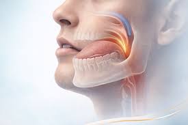
- Class II (Retrognathia – Mandibular Recession): The upper jaw is protruding or the lower jaw is recessed relative to the skull base. A “bird’s face” appearance may occur in profile. It is often associated with respiratory problems (sleep apnea).
- Class III (Prognathia): The lower jaw is significantly forward of the upper jaw. Chewing function is severely impaired, resulting in a “concave” profile.
- Apertognathia (Open Bite): This occurs when the posterior teeth meet but the anterior teeth do not. This creates difficulties in chewing and speaking.
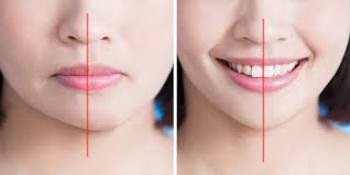
- Facial Asymmetry: This occurs when the jaw shifts to the right or left.
The Critical Role of Orthognathic Surgery in Treatment
The primary role of orthognathic surgery is to restore skeletal balance, beginning where orthodontics end. This role can be examined under three main headings:
A. Ensuring Occlusion
When there is a skeletal misalignment, it is physically impossible for the teeth to fully contact each other. Surgery moves the jaws into the ideal position, allowing the teeth to establish a proper lock-and-key relationship (intercuspation). This activates the chewing function, which is the beginning of the digestive system.
B. Increasing Airway Patency
In particular, in patients with a recessed lower jaw, the airway becomes narrower, increasing the risk of Obstructive Sleep Apnea Syndrome (OSAS). Maxillomandibular advancement (advancement) physically widens the airway, facilitating breathing.
C. Temporomandibular Joint (TMJ) Health
Malfunctions in occlusion can increase the load on the jaw joint. Surgery helps maintain joint health or manage existing symptoms by distributing unbalanced loads on the joint.
Diagnosis and Digital Planning Process
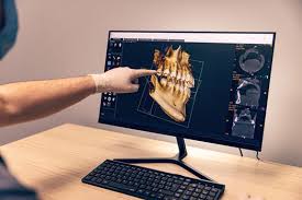
Modern orthognathic surgery is an engineering endeavor requiring millimeter precision. 3D digital planning has replaced traditional methods.
- Clinical Evaluation: Facial proportions, smile line, gingival appearance, and jaw movements are analyzed.
- Imaging: The patient’s head structure is transferred to a 3D virtual environment using panoramic x-rays, cephalometric analysis, and most importantly, Cone Beam Computed Tomography (CBCT).
- Virtual Surgical Simulation (VSP): The surgeon simulates the surgery in a computer environment. The extent of the bone to be cut and the angle at which it will be moved are calculated.
- Surgical Splint Production: 3D printers are used to produce devices (splints) that will guide the new position of the jaws during surgery, in accordance with the plan.
Surgical Techniques and Procedures Applied
Orthognathic surgery is performed under general anesthesia in fully equipped hospital settings. Because the incisions are made entirely inside the mouth, no visible scars are left on the face.
| Technique | Area of Application | Purpose |
|---|---|---|
| Le Fort I Osteotomy | Upper Jaw (Maxilla) | Moving the upper jaw forward, backward, up, or down. Used to treat a gummy smile or maxillary recession. |
| BSSO (Bilateral Sagittal Split Osteotomy) | Lower Jaw (Mandibula) | Moving the lower jaw forward or backward. Preserving the nerve tissue and positioning the jawbone in the sagittal plane.It is separated and shifted. |
| Genioplasty | Chin | Aesthetic and functional reshaping of the chin. Important for profile balance. |
| SARPE (Surgically Assisted Rapid Palatal Expansion) | Maximal Expansion | Expansion of the narrow upper jaw in adult patients with surgical assistance. |
Beyond Aesthetics: Functional Gains
Although the primary motivation for patients to choose this surgery is generally improvement in facial appearance, the true success of the surgery lies in functional improvements.
- Effective Chewing: Proper grinding of food reduces digestive system problems (reflux, indigestion).
- Speech (Articulation): The correct position of the teeth and jaw helps the tongue produce sounds correctly. Problems such as lisping can be corrected.
- Lip Closure: In patients with lip insufficiency, the mouth remains open at rest. After surgery, the lips close comfortably, preventing dry mouth and gum disease.
- Respiratory Quality: Airway expansion increases oxygen saturation and improves sleep quality.
Pre- and Post-Operative Management
Orthognathic surgery is a process consisting of the “Orthodontics – Surgery – Orthodontics” trilogy.
- Pre-Operative Orthodontics (Decompensation): Before surgery, braces are placed on the teeth. The goal is to align the teeth to their ideal angles within the jawbone. During this process, the patient’s appearance may temporarily appear more noticeable because the skeletal problem masked by the teeth is revealed.
- Surgery: Surgery is performed while the braces are in place. After surgery, the jaws are usually guided together with elastics.
- Post-Operative Orthodontics: After surgery, braces continue for another 6-9 months to fine-tune the bite and establish the occlusion.
Recovery Nutrition
A liquid diet is recommended for the first week after surgery, followed by soft foods (puree consistency) that do not require chewing in the following weeks. Bone healing (consolidation) takes approximately 6-8 weeks. Avoiding hard foods during this period is essential for the stability of the plate and screws.
Medical Risks and Complication Management
As with any surgical procedure, orthognathic surgery carries potential risks, and patients should be informed in detail about these risks.
- Paresthesia (Loss of Sensation): During lower jaw surgery, temporary loss of sensation at the tip of the lip and chin may occur due to manipulation of the inferior alveolar nerve. In most cases, it returns within 6 months; rarely, it can be permanent.
- Infection: The oral environment is rich in bacterial flora. This is managed with antibiotic use and oral hygiene.
- Relapse: This is the tendency for the jaw to return to its previous position. This risk is minimized with proper planning and rigid fixation (plate-screw system).
Conclusion
Orthognathic surgery in the treatment of jaw disorders is the intersection of science, technology, and art. It not only corrects facial proportions but also improves the patient’s most basic life functions, from breathing to eating. This multidisciplinary process, conducted in collaboration with an orthodontist and an oral surgeon, provides patients with a permanent and healthy chewing system that will last a lifetime.