What is Orthognathic Surgery? Medical Definition
Orthognathic surgery is derived from the Greek words “orthos” (straight) and “gnathos” (jaw). In medical literature, it is defined as the surgical correction of skeletal irregularities in the lower jaw (mandible), upper jaw (maxilla), and facial bones.
This procedure is used when orthodontic treatment (braces), which only straighten the teeth, is inadequate. Even if the teeth are aligned properly, if the position, size, or relationship of the jawbones is misaligned, orthodontic treatment alone cannot provide a functional occlusion. Orthognathic surgery repositions the jaws, achieving both functional (chewing, breathing) and aesthetic (facial profile) harmony.
Who is Suitable for Orthognathic Surgery?
Not every jaw disorder requires surgery. However, surgery is unavoidable in cases where the extent of skeletal deviation exceeds certain limits. Ideal candidates generally possess the following characteristics:
- Completion of Growth and Development: Surgery is usually performed after skeletal growth has stopped. This is usually around the age of 17-18 for women and 18-21 for men. Early surgery may lead to a risk of recurrence (recurrence of the problem) due to continued growth.
- Skeletal Discrepancy: Individuals whose radiographic analysis has proven that the problem is not solely dental but skeletal.
- Systemic Health Status: Individuals who do not have a serious systemic condition that would prevent them from undergoing general anesthesia or major surgery.
- Realistic Expectations: Patients who are aware that treatment is a long-term process (orthodontics + surgery) and who can adapt to it.
Basic Symptoms and Clinical Indications
Symptoms observed in patients requiring orthognathic surgery are functional problems that directly affect their quality of daily life. If one or more of the following symptoms are present, a consultation with an Oral and Maxillofacial Surgeon or Orthodontist is necessary:
Functional Symptoms
- Chewing and Biting Difficulty: Inability to tear food with the front teeth or grind it completely with the back teeth.
- Swallowing Problems: Difficulty swallowing due to improper tongue positioning.
- Chronic Mouth Breathing: Inability to breathe through the nose and constant mouth breathing due to the narrowing of the airway by the jaw structure.
- Sleep Apnea (OSAS): Cessation of breathing during sleep, snoring, and poor quality sleep. It is particularly common in cases where the lower jaw is retracted.
- TMJ (Jaw Joint) Pain: Chronic pain, audible sound, or limitation in the jaw joint due to malocclusion.
- Speech Disorders: Difficulty pronouncing certain letters (such as s, ş, z).
Aesthetic and Physical Symptoms
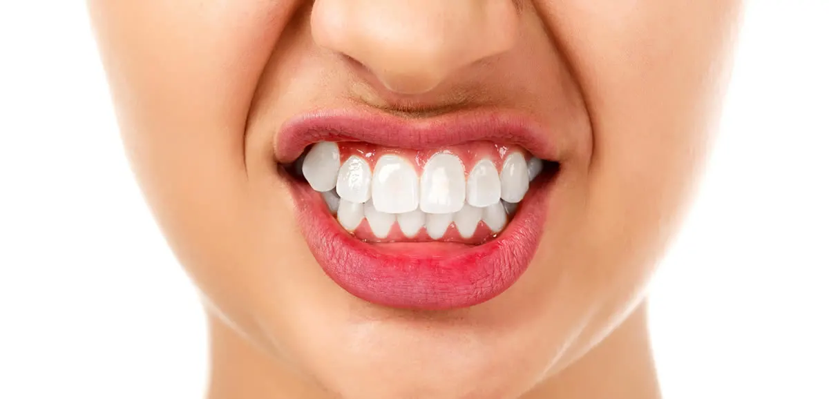
- The jaw is positioned forward (prognathism) or backward (retrognatism).
- Significant facial asymmetry (jaw shifting to one side).
- Incompetent lips.
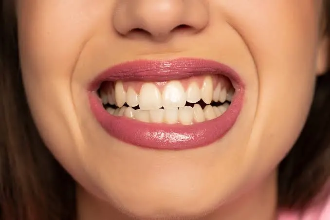
- Excessive gum display when smiling (Gummy Smile).
- The jaw appears too long or too short.
Specific Skeletal Deformities Corrected
Anomalies corrected with orthognathic surgery are categorized according to the Angle Classification and skeletal analyses.
| Anomaly Type | Definition | Surgical Target | |
|---|---|---|---|
| Class II Malocclusion (Retrognathia) | Excessive retraction of the lower jaw relative to the upper jaw (“Bird face” appearance). | Forward displacement of the lower jaw (BSSO) or retraction of the upper jaw. | |
| Class III Malocclusion (Prognathia) | Excessive protraction of the lower jaw relative to the upper jaw. | Retraction of the lower jaw and/or protraction of the upper jaw Removal. | |
| Apertognathia (Open Bite) | The anterior teeth do not touch each other while the posterior teeth touch, leaving a gap. | The anterior bite is achieved by impaction of the posterior part of the upper jaw. | |
| Facial Asymmetry | The jaw shifts to the right or left, deviation of the facial midline. | Achieving symmetry by bringing the jaws into the midline. | |
| Vertical Maxillary Excess (Gummy Smile) | Vertical maxillary excess (Gummy Smile) | Excessive vertical growth of the upper jaw, excessive gingival display during smile. | The upper jaw is positioned upwards (Le Fort I impaction). |
Diagnosis and Planning Process: Digital Transformation
The success of orthognathic surgery depends on planning with millimeter precision. Traditional methods have now been replaced by 3D digital planning. The diagnostic process includes the following steps:
Clinical Examination
The patient’s facial proportions, dental occlusion, joint health, and respiratory functions are physically evaluated. The patient’s complaints and expectations are listened to.
Radiologic Imaging
- Panoramic X-ray: The general condition of all teeth and jawbones is visualized.
- Cephalometric X-ray: The angular relationships between the skull bones and jaws are analyzed.
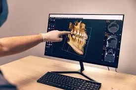
- CBCT (Cone Beam Computed Tomography): This is the most critical stage. An exact 3D model of the patient’s skull is created. Nerve locations, root positions, and bone thickness are examined in 3D.
Digital Planning and Simulation
CBCT data is transferred to specialized software. The surgeon performs the surgery in a virtual environment (Virtual Surgical Planning – VSP). The extent of jaw movement and the location of the incisions are calculated to the nearest millimeter. During this phase, the patient can see their projected post-operative facial profile. Based on this planning, the “Surgical Guides” (Splints) to be used during the surgery are produced with 3D printers.
Treatment Journey: Orthodontics and Surgical Collaboration
Orthognathic surgery is not a stand-alone procedure; it is an integrated “Teamwork” with orthodontics. The process generally consists of three main phases.
Phase 1: Preoperative Orthodontics (Decompensation)
Teeth may naturally be misaligned to mask the skeletal deformity (Dental Compensation). Preoperative braces are used to adjust the teeth to their ideal angles within the jaw. During this phase, the patient’s appearance may temporarily appear “worse” as the masking is removed and the true skeletal problem is revealed. This process takes an average of 6-12 months.
Phase 2: Surgery
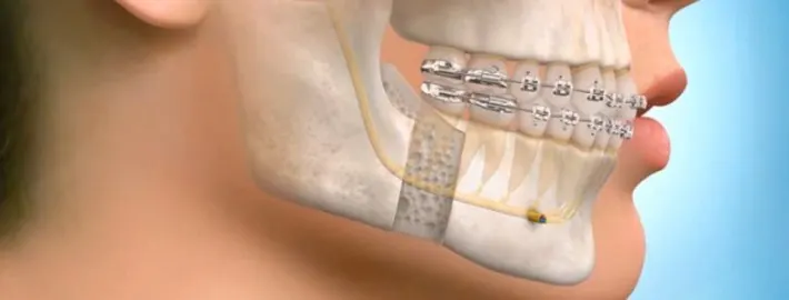
Surgery is performed once the teeth are ready. The patient enters the surgery with braces.
Phase 3: Postoperative Orthodontics
After surgery, orthodontic treatment continues for another 4-6 months to allow the teeth to fully align (fine-tune) in the new jaw position. Finally, the braces are removed and retention treatment is initiated.
Note: In some suitable cases, a “Surgery First” approach may be applied. In this method, surgery is performed first, then the wires are attached. This decision is made by the physician.
Surgical Techniques Applied
Orthognathic surgery is performed under general anesthesia in a fully equipped hospital setting. The most commonly performed procedures are:
Le Fort I Osteotomy (Upper Jaw Surgery)
The upper jawbone (maxilla) is cut horizontally at the level of the base of the nose. After the upper jaw is freed, it is moved to its new planned position (forward, backward, upward, or downward). It is fixed in the new position with titanium plates and screws. The incisions are made entirely intraorally, leaving no facial scars.
BSSO (Bilateral Sagittal Split Osteotomy – Lower Jaw Surgery)
The lower jawbone (mandible) is cut bilaterally from the posterior region (ramus) using a special technique and separated like two leaves. The chin is shifted forward or backward, preserving the nerve tissue. It is fixed with plates and screws.
Genioplasty (Chin Tip Aesthetics)
The chin can be cut independently of the rest of the lower jaw and moved forward, backward, or sideways. It is an important touch that completes the facial profile.
Recovery Process and Post-Operative Care
The recovery process varies depending on the patient’s age and the extent of the surgery, but a general timetable is as follows:
- Hospitalization: Usually 1-3 days.
- First Week: Facial edema (swelling) and bruising occur. This peaks in days 3-4 and then begins to subside. Pain management is provided with medications.
- Diet: A completely liquid diet is followed for the first week. Because the jaws may be restrained with elastics, mouth opening is limited. Soft foods (purées) are introduced starting in the second week. A full return to chewing function usually takes 4-6 weeks.
- Hygiene: Oral hygiene is critical because there are incisions in the mouth. A physician-recommended mouthwash and gentle brushing techniques should be used.
- Return to Work/School: 2-3 weeks of rest is generally recommended.
- Complete Bone Union: takes 6-8 weeks. Protection from impacts is essential during this period.
Possible Risks and Complication Management
As with any surgical procedure, orthognathic surgery carries risks. Possible complications based on scientific data:
- Nerve Damage (Paresthesia): AbstractTemporary loss of sensation or tingling may occur at the tip of the lip and chin, especially during lower jaw surgery. In most cases, this resolves within 6 months; rarely, it can be permanent.
- Infection: There is a risk of infection due to oral flora, which is managed with antibiotics.
- Relapse: This is the tendency for the jaw to return to its previous position. This risk is minimized with proper planning and rigid fixation (plates and screws).
- TMJ Problems: Rarely, new problems may develop in the jaw joint, or existing problems may change.
Frequently Asked Questions
Will the surgery leave a scar?
No, orthognathic surgery incisions are made entirely inside the mouth. There are no visible scars on the face. If only the tip of the jaw or very small holes (for screw placement) are required, temporary scars of 2-3 mm may be possible.
How long will my jaws be fixed?
In the past, jaws were tightly fixed with wires for 6-8 weeks. Today, because “rigid fixation” (titanium plates and screws) is used, complete jaw fixation is rare. Usually, only guiding elastics are used, and the patient can open their mouth, albeit limitedly.
Is the surgery painful?
Patients generally complain of swelling (edema) and a feeling of tightness after surgery, rather than pain. Due to the temporary nerve damage, the pain is less severe than expected. Painkillers provide a comfortable experience.
Will my speech be affected?
Speaking may be somewhat difficult in the first weeks due to the swelling and elastics. However, once healing is complete, speech and articulation (pronouncing letters) are usually much better than before, as the skeletal structure improves.