Tooth Extraction Application Methods in Kahramanmaraş: Anatomy-Specific Surgical Engineering at Dentocare Clinic
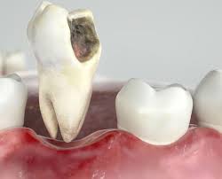
In dental practice, although the removal of a tooth from the jawbone may appear from the outside to be a uniform procedure, each patient’s bone density, root anatomy, and the sensitivity of the surrounding tissues are as unique as a fingerprint. These anatomical differences make a standard, memorized extraction procedure impossible. Safely removing a decayed, broken, or deeply impacted tooth that remains embedded in the jawbone at an incorrect angle is only possible through tooth extraction application methods that are specially modified for the case and require advanced surgical experience. Dentocare Clinic, serving the Kahramanmaraş region with advanced surgical standards, does not impose a “one-size-fits-all” extraction on its patients. Instead, it implements personalized surgical application methods that are best suited to the biological character of the problem, preserve the jawbone to the maximum extent, and shorten the healing process.
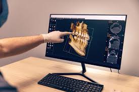
In traditional dentistry, the philosophy of “pulling the tooth out with brute force” has long been replaced by modern application methods in which technique and biomechanics speak louder than force. Before the operation, Dentocare Clinic’s specialist maxillofacial surgeons create a three-dimensional digital map of your jawbone using Dental Tomography (CBCT), planning well in advance the angle at which the tooth will be approached, which bone wall will be flexed, and—if necessary—at which points the tooth will be sectioned into parts before removal. Every method applied in our clinic is built upon the principle of “causing zero harm to the bone and tissue left behind while removing the tooth” (atraumatic surgery). Now, let us take a closer look at the details of these advanced application methods that require technology and expertise.
Elegance Replacing Force: The Atraumatic Luxation (Flexing) Technique
For standard teeth that have fully erupted into the mouth, whose crown (upper part) is intact, and whose roots are not excessively hook-shaped, our clinic’s first choice is always the Atraumatic Luxation Technique. The main aim of this method is to release the tooth from its socket without fracturing the thin surrounding bone walls (alveolar bone) and without tearing the gum tissue. In older methods, the tooth would be directly gripped with forceps and pulled. In this modern technique, however, the process follows a far more delicate biomechanical sequence.
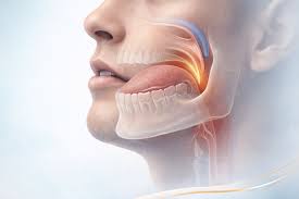
The procedure begins with syndesmotomy, in which the connection between the tooth and the gum is delicately severed with micro-scalpels. Then, using fine-tipped surgical elevators, the dentist enters millimetrically between the tooth and the bone. The tooth is flexed very gently within its socket with vibratory movements to the right, left, and upward. This luxation movement tires and separates the microscopic fibers (periodontal ligaments) that anchor the tooth to the bone. Once the tooth feels as though it has “fallen loose” inside the bone, it is removed gently without applying any force. This method, which does not require sutures, is the most comfortable application technique and greatly minimizes post-extraction pain and swelling.
Odontosection for Difficult Cases: Sectional Tooth Removal
One of the anatomical obstacles that frightens patients most during tooth extraction is having a tooth with multiple roots that diverge in opposite directions within the jawbone, much like the roots of a tree. If a dentist attempts to remove such a tooth in one single piece, those opposing roots exert immense pressure on the surrounding bone from within, potentially causing cracks or even major fractures of the jawbone. At Dentocare Clinic, this anatomical risk is completely eliminated through the Odontosection (Sectional Tooth Removal) Method.
In the odontosection method, instead of forcing the entire tooth out as one unit, our dentist uses special physiodyspenser devices (surgically cooled motors) to divide the tooth into two or three parts while it is still inside the mouth. Once the tooth loses its structural integrity and resistance, each root fragment can be removed individually within seconds, following its own خروج angle without catching on the surrounding bone. Although the idea of breaking the tooth into pieces may sound intimidating psychologically, odontosection is in fact the art of sacrificing the tooth in order to protect the bone. This application is a true engineering marvel, especially in lower molars, where it reduces surgical trauma to nearly zero.
Comparison of Surgical Methods by Anatomical Challenge
Which tooth is extracted with which method is determined according to the anatomical condition of the tooth. In the table below, you can examine the differences between traditional methods and Dentocare Clinic’s advanced surgical applications used when standard approaches are insufficient:
| Clinical Condition (Anatomical Obstacle) | Old-Type / Standard Application | Dentocare Advanced Application Method |
|---|---|---|
| Roots Broken Below the Jawbone | The root is searched for blindly with instruments, causing crushing of the surrounding bone and major gum tears. | Flap Operation: The gum is opened over the bone with an aesthetic incision, the root is removed under direct vision and safely, and the area is closed with micro-sutures. |
| Deeply Impacted Wisdom Teeth Close to the Nerve | When bone is removed with rotary burs, there is a high risk of damaging the inferior alveolar nerve. | Piezosurgery Application: The bone is cut using ultrasonic sound waves that do not damage the nerve, reducing the risk of nerve injury to zero. |
| Teeth Fused with the Bone (Ankylosis) | Excessive force is applied because the tooth is fused with the bone, which may cause trauma to the temporomandibular joint (TMJ). | Combined Approach: The bone is protected by applying odontosection and minimal osteotomy with zero pressure on the jawbone. |
Piezosurgery and Osteotomy Applications for Impacted Teeth
Another application considered a technological revolution among tooth extraction methods is Piezosurgery-Assisted Osteotomy. In order to reach wisdom teeth that remain horizontally or inversely impacted inside the jawbone, a certain amount of bone tissue covering the tooth must be removed. In old-style methods, this was done with high-speed burs that produced heat, often resulting in bruising and serious swelling in the cheeks after surgery.
At Dentocare Clinic, the Piezosurgery method uses three-dimensional ultrasonic vibrations rather than mechanical friction to cut bone. The greatest feature of this remarkable device is its ability to perform selective cutting: it cuts only hard tissue (bone) and does not cut soft tissue even if the tip accidentally touches a blood vessel or nerve. Since bone cells are not burned during tooth extraction procedures performed with piezosurgery, our patients experience almost no pain after surgery and return to their social lives much more quickly.
The Final Step of the Application: Closure of the Biological Field (Suturation)
A surgical tooth extraction is not completed when the tooth is removed, but when the area is closed in the way most suitable for the body’s healing system. After extractions requiring advanced surgery (flap or osteotomy), the open tissue is not closed with an ordinary stitch, but with Micro-Suturation Methods applied with the delicacy of plastic surgery.
Dentocare Clinic surgeons use medical sutures fine enough not to disrupt blood circulation (ischemia), bringing the wound edges together without tension. In cases where it is necessary—especially when an implant is planned in the future—PRF (platelet-rich fibrin) obtained from the patient’s own blood or biocompatible bone graft material is placed into the socket before suturing (Socket Preservation). This is the final stage of engineering that stops jawbone resorption and transforms the extraction socket into strong bone tissue over the coming months.
The Dentocare Difference for Anatomy-Specific Surgical Applications
Tooth extraction is not a factory-made procedure in which the same instrument can be used in the same way for every patient. A successful and complication-free surgical process requires a mind capable of correctly reading your jaw tomography, state-of-the-art devices such as piezosurgery, and experienced surgeons who translate the philosophy of “protecting the bone” into manual practice. Old-style forceful applications may cause bone loss and nerve damage that can cost tens of thousands later to repair.
If you have been told in Kahramanmaraş and the surrounding area that “this tooth will be very difficult to extract, it is embedded in the bone, or the root is broken,” and you feel worried about the surgical process, it is time to discover Dentocare Clinic’s modern application methods. Dentocare Clinic transforms even the most difficult anatomical obstacles into a safe and comfortable healthcare step through advanced surgical methods such as atraumatic luxation, odontosection, and piezosurgery. To determine the most suitable and most comfortable surgical application method for your own anatomy, you can contact our clinic immediately and schedule your detailed radiological analysis appointment today.